Copyright
©The Author(s) 2022.
World J Gastroenterol. Jun 21, 2022; 28(23): 2569-2581
Published online Jun 21, 2022. doi: 10.3748/wjg.v28.i23.2569
Published online Jun 21, 2022. doi: 10.3748/wjg.v28.i23.2569
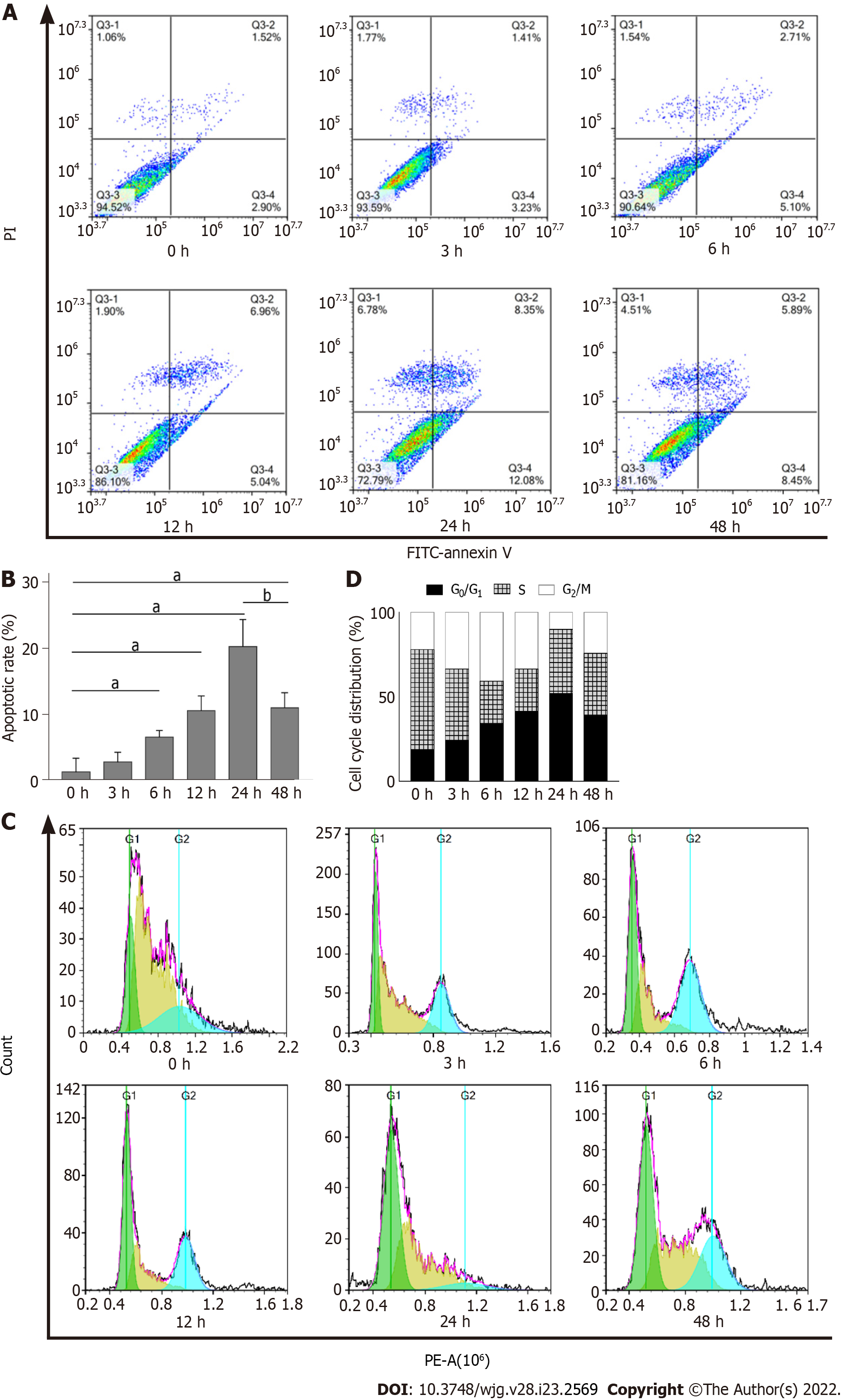
Figure 4 Impact of dithiothreitol treatment on cell cycle and apoptosis of buffalo rat liver 3A cells.
A and B: Buffalo rat liver 3A (BRL-3A) cells were treated with 2.0 mmol/L dithiothreitol (DTT) for 0, 3, 6, 12, 24 and 48 h. The population of apoptotic cells was detected by flow cytometry. The lower right quadrant represents the early apoptotic cells, and the upper right quadrant represents the late apoptotic cells; C and D: BRL-3A cells were treated with 2.0 mmol/L DTT for 0, 3, 6, 12, 24 and 48 h. The analysis of the cell cycle was assessed by flow cytometry. aP < 0.05 vs 0 h group; bP < 0.05 vs 48 h group.
- Citation: Guo YX, Han B, Yang T, Chen YS, Yang Y, Li JY, Yang Q, Xie RJ. Family with sequence similarity 134 member B-mediated reticulophagy ameliorates hepatocyte apoptosis induced by dithiothreitol. World J Gastroenterol 2022; 28(23): 2569-2581
- URL: https://www.wjgnet.com/1007-9327/full/v28/i23/2569.htm
- DOI: https://dx.doi.org/10.3748/wjg.v28.i23.2569


