Copyright
©The Author(s) 2022.
World J Gastroenterol. Jun 14, 2022; 28(22): 2482-2493
Published online Jun 14, 2022. doi: 10.3748/wjg.v28.i22.2482
Published online Jun 14, 2022. doi: 10.3748/wjg.v28.i22.2482
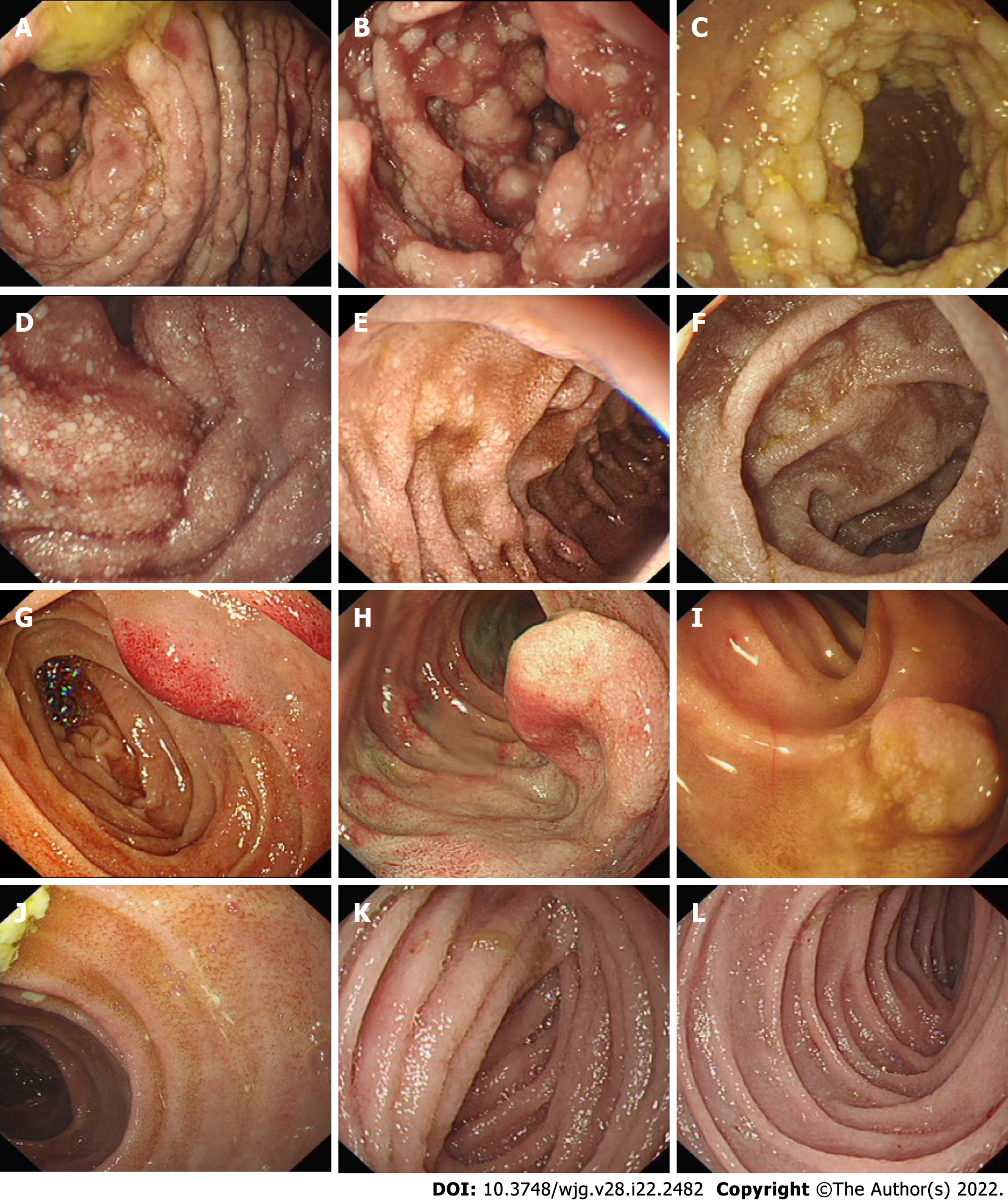
Figure 2 Endoscopic images of patients with primary intestinal lymphangiectasia.
The images show white mucosal plaques and spots in the small intestine, divided into four types. A-C: Nodular type; D-F: Granular type; G-I: Vesicular type; J-L: Edematous type.
- Citation: Meng MM, Liu KL, Xue XY, Hao K, Dong J, Yu CK, Liu H, Wang CH, Su H, Lin W, Jiang GJ, Wei N, Wang RG, Shen WB, Wu J. Endoscopic classification and pathological features of primary intestinal lymphangiectasia. World J Gastroenterol 2022; 28(22): 2482-2493
- URL: https://www.wjgnet.com/1007-9327/full/v28/i22/2482.htm
- DOI: https://dx.doi.org/10.3748/wjg.v28.i22.2482


