Copyright
©The Author(s) 2022.
World J Gastroenterol. Jun 7, 2022; 28(21): 2396-2402
Published online Jun 7, 2022. doi: 10.3748/wjg.v28.i21.2396
Published online Jun 7, 2022. doi: 10.3748/wjg.v28.i21.2396
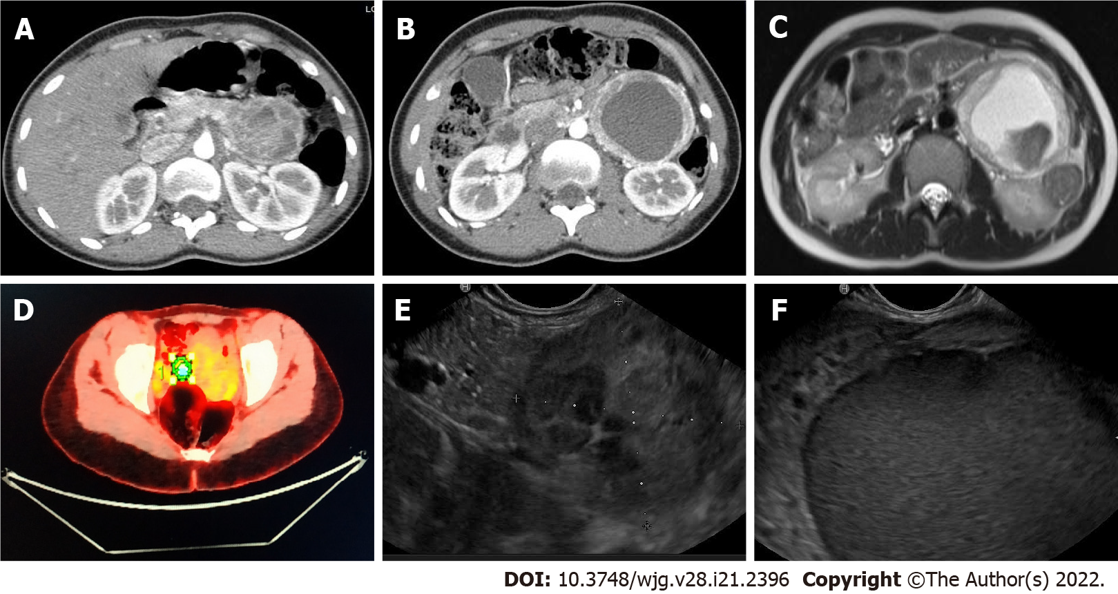
Figure 1 Radiologic findings.
A and B: Computed tomography-scan showed a 95-mm cystic lesion with no cleavage plane from the pancreas; C: Nuclear magnetic resonance evidenced that the lesion was hyper-intense in T1; D: Positron emission tomography demonstrated a 4.7% standard uptake value; E and F: Endoscopic ultrasonography identified a hypoechoic mass close to the pancreatic tail.
- Citation: Petrelli F, Fratini G, Sbrozzi-Vanni A, Giusti A, Manta R, Vignali C, Nesi G, Amorosi A, Cavazzana A, Arganini M, Ambrosio MR. Peripancreatic paraganglioma: Lesson from a round table. World J Gastroenterol 2022; 28(21): 2396-2402
- URL: https://www.wjgnet.com/1007-9327/full/v28/i21/2396.htm
- DOI: https://dx.doi.org/10.3748/wjg.v28.i21.2396


