Copyright
©The Author(s) 2022.
World J Gastroenterol. May 21, 2022; 28(19): 2112-2122
Published online May 21, 2022. doi: 10.3748/wjg.v28.i19.2112
Published online May 21, 2022. doi: 10.3748/wjg.v28.i19.2112
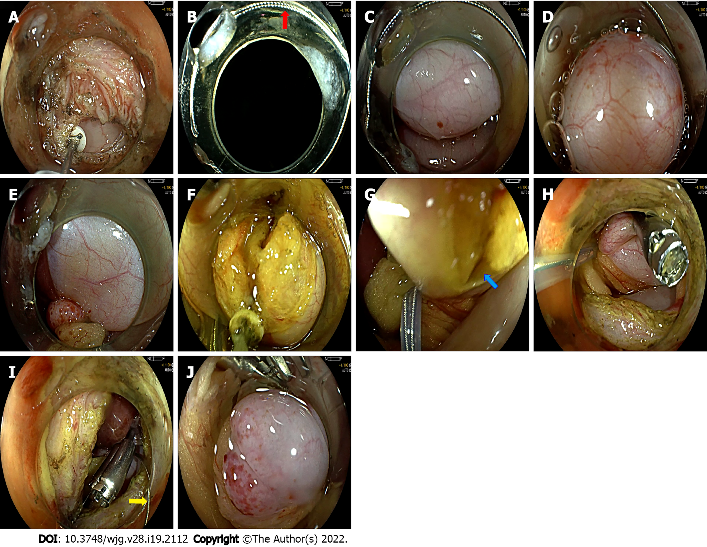
Figure 1 Snare-assisted flexible endoscope in transgastric endoscopic gallbladder-preserving surgery.
A: The anterior wall of the gastric antrum was incised; B: The snare was placed on the transparent cap (red arrow); C: The endoscope was inserted into the abdominal cavity. The gallbladder was located; D: The transparent cap clung to the gallbladder wall, and the gallbladder wall was sucked in it. The snare was released, and the gallbladder wall was ligated; E: This ligation state was maintained; F: The gallbladder wall was incised with the assistance of the snare; G: With the navigation of the snare, the endoscopist quickly found the gallbladder and gallbladder incision again (blue arrow); H: With the help of the snare, clips were used to close the gallbladder wall incision; I: The snare was loosened (yellow array); J: The gallbladder wall returned to normal.
- Citation: Guo XW, Liang YX, Huang PY, Liang LX, Zeng YQ, Ding Z. Snare-assisted flexible endoscope in trans-gastric endoscopic gallbladder-preserving surgery: A pilot animal study. World J Gastroenterol 2022; 28(19): 2112-2122
- URL: https://www.wjgnet.com/1007-9327/full/v28/i19/2112.htm
- DOI: https://dx.doi.org/10.3748/wjg.v28.i19.2112


