Copyright
©The Author(s) 2022.
World J Gastroenterol. May 21, 2022; 28(19): 2100-2111
Published online May 21, 2022. doi: 10.3748/wjg.v28.i19.2100
Published online May 21, 2022. doi: 10.3748/wjg.v28.i19.2100
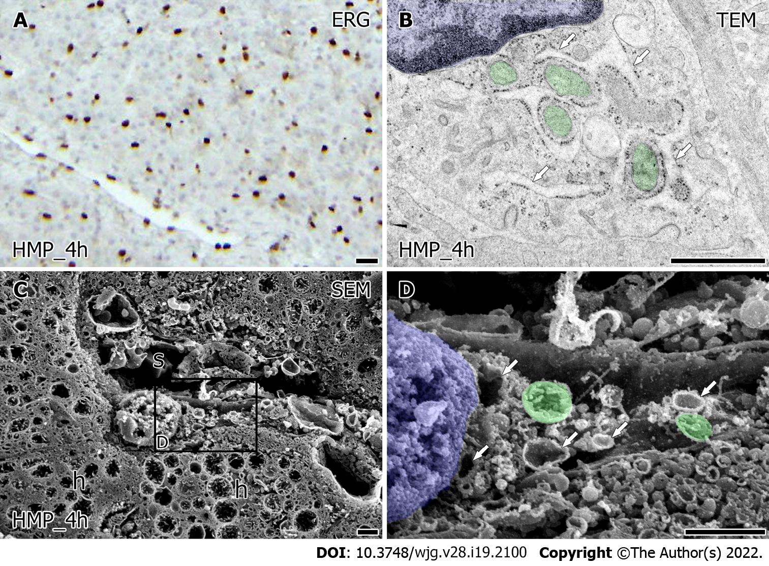
Figure 4 The ultrastructural alteration in porcine liver sinusoidal endothelial cells preserved by hypothermic machine perfusion.
A: The distribution of viable liver sinusoidal endothelial cells (LSEC) which is indicated with ERG-positive cells in the porcine liver preserved by hypothermic machine perfusion (HMP) for 4 h after 60 min of warm ischemia. Bar = 10 μm; B: transmission electron microscopy image of the LSEC in porcine liver preserved by HMP for 4 h after 60 min of warm ischemia. Bars = 1 μm; C and D: LSEC was observed by scanning electron microscopy in osmium-macerated porcine liver preserved by HMP for 4 h after 60 min of warm ischemia. s: Sinusoid. h: Hepatocyte. The partial area indicated in C was further observed in high magnification D. Bars = 1 μm. Mitochondria are labeled in green and the nucleus is labeled in blue. Arrows indicate the rER. TEM: Transmission electron microscopy; SEM: Scanning electron microscopy.
- Citation: Bochimoto H, Ishihara Y, Mohd Zin NK, Iwata H, Kondoh D, Obara H, Matsuno N. Ultrastructural changes in porcine liver sinusoidal endothelial cells of machine perfused liver donated after cardiac death. World J Gastroenterol 2022; 28(19): 2100-2111
- URL: https://www.wjgnet.com/1007-9327/full/v28/i19/2100.htm
- DOI: https://dx.doi.org/10.3748/wjg.v28.i19.2100


