Copyright
©The Author(s) 2022.
World J Gastroenterol. May 7, 2022; 28(17): 1814-1829
Published online May 7, 2022. doi: 10.3748/wjg.v28.i17.1814
Published online May 7, 2022. doi: 10.3748/wjg.v28.i17.1814
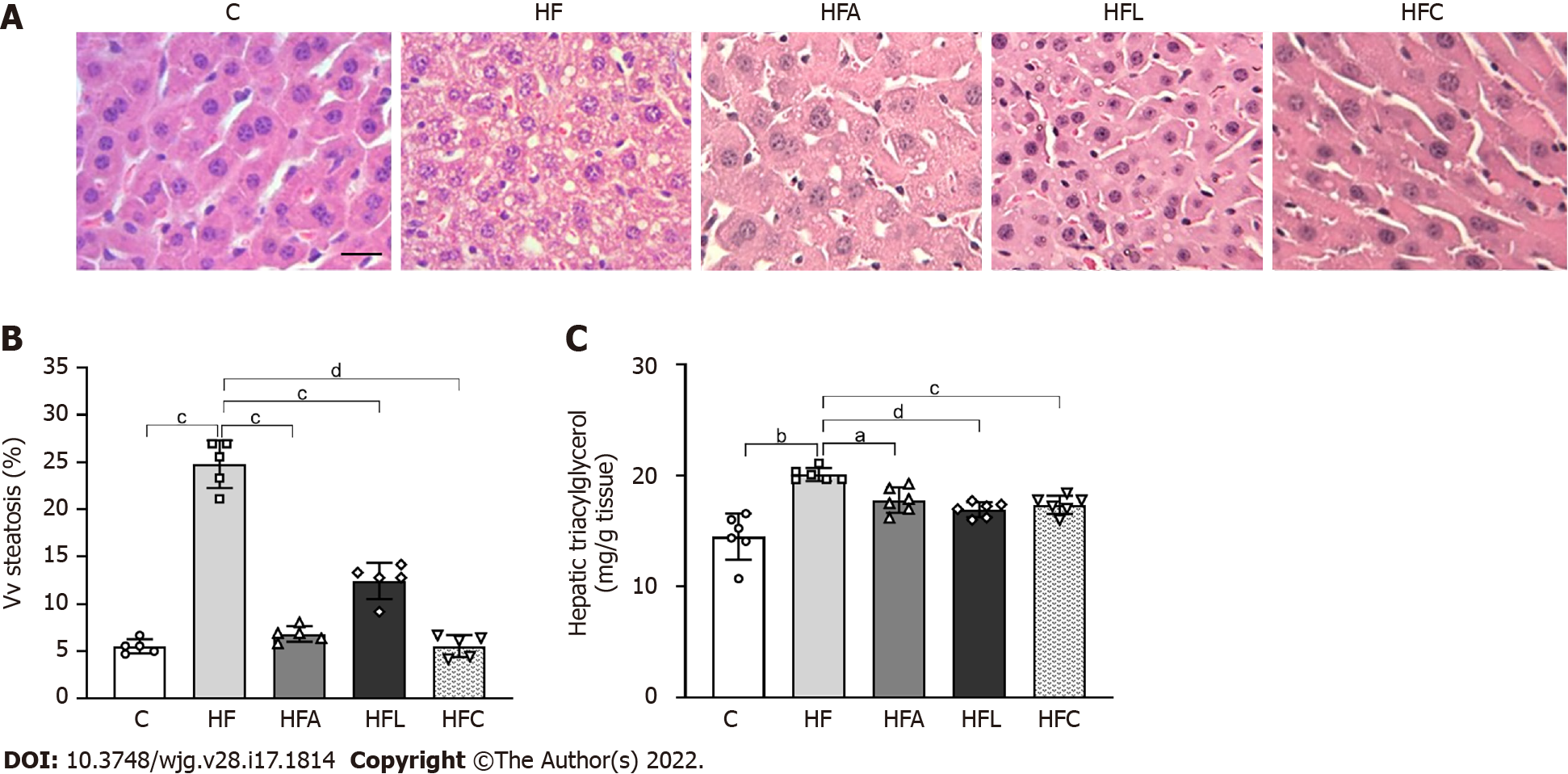
Figure 5 Liver histology, stereology, and biochemistry.
A: Hematoxylin-eosin liver sections; B: Volume density (Vv) (liver steatosis); C: Hepatic triacylglycerol. Liver sections show widespread hepatic steatosis after chronic HF diet intake and expressive reduction in all treated groups (scale bar = 50 μm. Both stereology (Vv steatosis) and biochemical analyses (hepatic triacylglycerol) confirm these observations. Brown-Forsythe and Welch one-way ANOVA and Dunnett T3 post hoc test (mean ± SD, n = 6 for biochemistry and n = 5 for stereology). Significant differences are indicated as follows: aP < 0.05; bP < 0.01; cP < 0.001; dP < 0.0001. C: Control diet; HF: High-fat diet; HFA: High-fat diet plus PPAR-alpha agonist (WY14643); HFL: High-fat diet plus DPP-4 inhibitor (linagliptin); HFC: High-fat diet plus the combination of WY14643 with linagliptin; Vv: Volume density.
- Citation: Silva-Veiga FM, Miranda CS, Vasques-Monteiro IML, Souza-Tavares H, Martins FF, Daleprane JB, Souza-Mello V. Peroxisome proliferator-activated receptor-alpha activation and dipeptidyl peptidase-4 inhibition target dysbiosis to treat fatty liver in obese mice . World J Gastroenterol 2022; 28(17): 1814-1829
- URL: https://www.wjgnet.com/1007-9327/full/v28/i17/1814.htm
- DOI: https://dx.doi.org/10.3748/wjg.v28.i17.1814


