Copyright
©The Author(s) 2022.
World J Gastroenterol. May 7, 2022; 28(17): 1768-1780
Published online May 7, 2022. doi: 10.3748/wjg.v28.i17.1768
Published online May 7, 2022. doi: 10.3748/wjg.v28.i17.1768
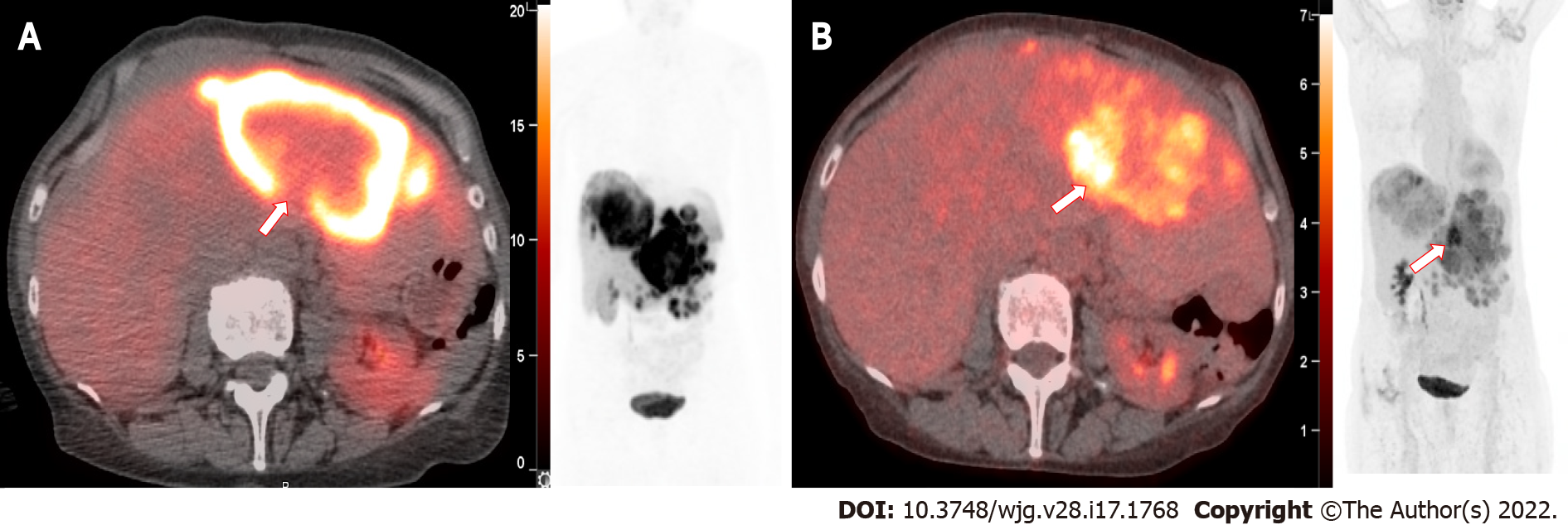
Figure 3 73 year old female with well-differentiated neuroendocrine tumor (Ki-67 = 6%) and liver metastases with an unknown primary.
A: 68Ga DOTATATE positron emission tomography (PET)/computed tomography (CT) fusion and whole-body PET projection confirms multifocal hepatic metastases SUVmax 34.4 with an area of focal decreased DOTATATE avidity (arrow); B: Fluorodeoxyglucose (FDG) PET/CT fusion and whole-body PET shows mismatched focal intense increased FDG uptake SUVmax 9.1 (arrow), suggestive of heterogeneous tumor phenotype with areas of dedifferentiation.
- Citation: Grey N, Silosky M, Lieu CH, Chin BB. Current status and future of targeted peptide receptor radionuclide positron emission tomography imaging and therapy of gastroenteropancreatic-neuroendocrine tumors. World J Gastroenterol 2022; 28(17): 1768-1780
- URL: https://www.wjgnet.com/1007-9327/full/v28/i17/1768.htm
- DOI: https://dx.doi.org/10.3748/wjg.v28.i17.1768


