Copyright
©The Author(s) 2022.
World J Gastroenterol. May 7, 2022; 28(17): 1751-1767
Published online May 7, 2022. doi: 10.3748/wjg.v28.i17.1751
Published online May 7, 2022. doi: 10.3748/wjg.v28.i17.1751
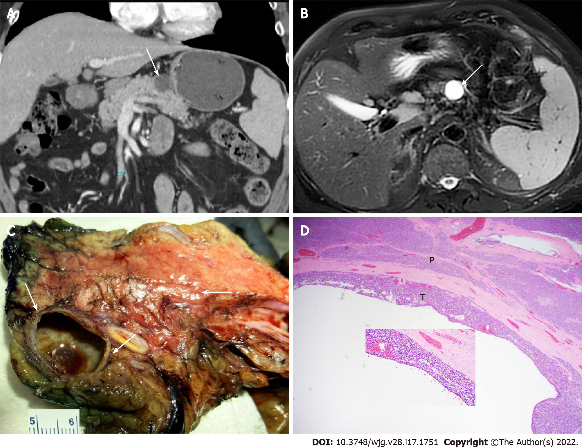
Figure 5 Pancreatic cystic neuroendocrine tumor mimicking mucinous cystic neoplasm.
A: Computed tomography image (axial) showing a 2.2 cm cystic lesion (arrow) in the pancreatic body of a 68-year-old man; B: Magnetic resonance imaging (T2, axial) showing the cystic mass (arrow); C: Partial pancreatectomy specimen showing cut surface of the thin-walled cystic pancreatic neuroendocrine tumor mimicking mucinous cystic neoplasm with no communication with the pancreatic duct (arrows); D: Histopathologic features of cystic pancreatic neuroendocrine tumor with acinar and trabecular architecture (T) and surrounding pancreatic parenchyma (P) (H&E stain, 40 ×, inset 200 ×).
- Citation: Yin F, Wu ZH, Lai JP. New insights in diagnosis and treatment of gastroenteropancreatic neuroendocrine neoplasms. World J Gastroenterol 2022; 28(17): 1751-1767
- URL: https://www.wjgnet.com/1007-9327/full/v28/i17/1751.htm
- DOI: https://dx.doi.org/10.3748/wjg.v28.i17.1751


