Copyright
©The Author(s) 2022.
World J Gastroenterol. May 7, 2022; 28(17): 1751-1767
Published online May 7, 2022. doi: 10.3748/wjg.v28.i17.1751
Published online May 7, 2022. doi: 10.3748/wjg.v28.i17.1751
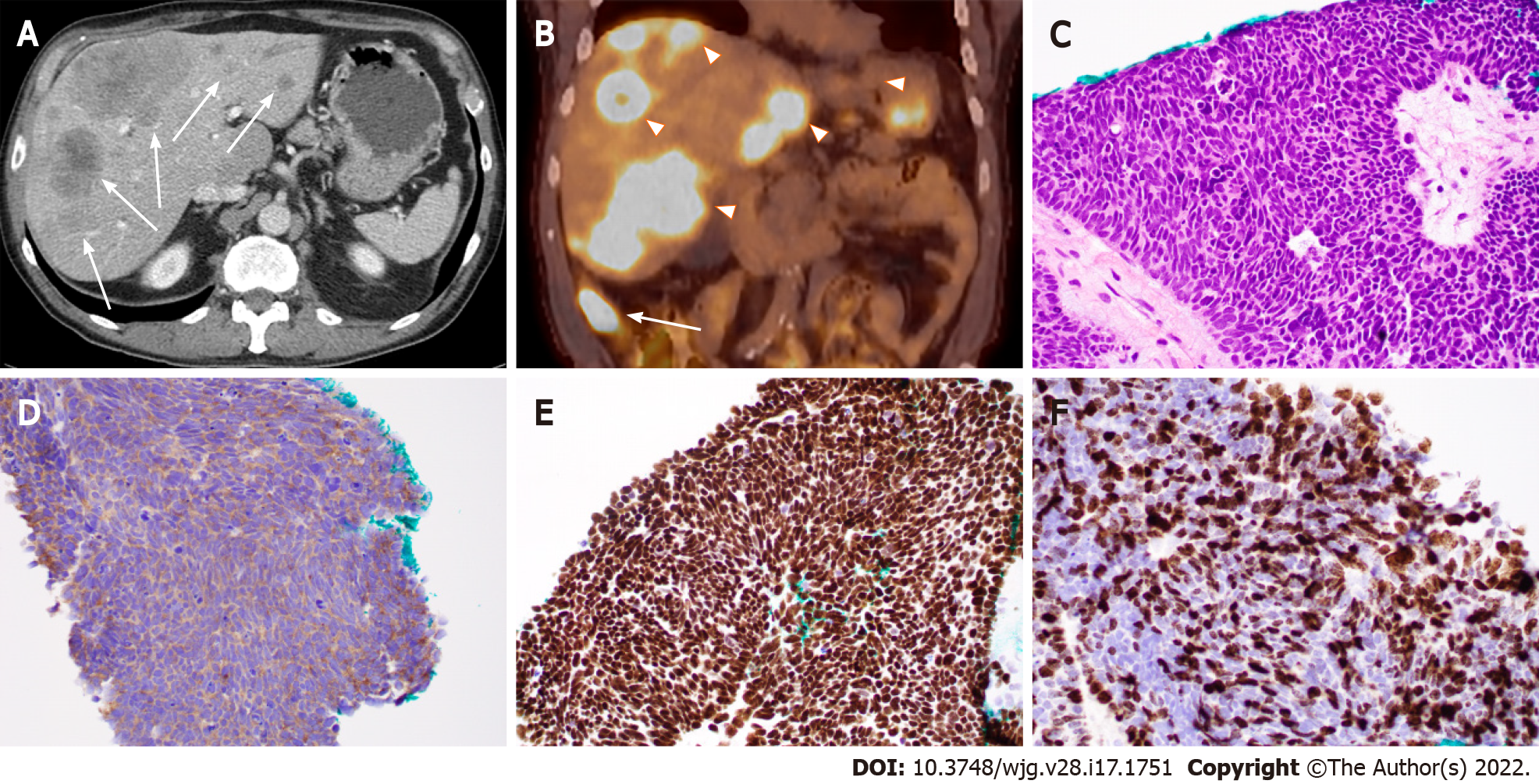
Figure 1 Ileal small cell neuroendocrine carcinoma with liver metastases in a 71-year-old man.
A: Computed tomography (CT) image (axial) showing ileal and multifocal liver lesions (arrow); B: Positron emission tomography-CT image showing ileal (arrow) and liver lesions (arrowheads) with hypermetabolic activity; C: Histopathologic features of small cell neuroendocrine carcinoma. Note the tumor cells with peripheral palisading, rosetting, scant cytoplasm, nuclear molding, finely granular chromatin, and lack of prominent nucleoli; D-F: Tumor cells with positive immunoreactivity for synaptophysin (D) and CDX2 (E) as well as a high Ki-67 proliferation index (70%) (F). (C-F: 400 ×).
- Citation: Yin F, Wu ZH, Lai JP. New insights in diagnosis and treatment of gastroenteropancreatic neuroendocrine neoplasms. World J Gastroenterol 2022; 28(17): 1751-1767
- URL: https://www.wjgnet.com/1007-9327/full/v28/i17/1751.htm
- DOI: https://dx.doi.org/10.3748/wjg.v28.i17.1751


