Copyright
©The Author(s) 2022.
World J Gastroenterol. Apr 28, 2022; 28(16): 1625-1640
Published online Apr 28, 2022. doi: 10.3748/wjg.v28.i16.1625
Published online Apr 28, 2022. doi: 10.3748/wjg.v28.i16.1625
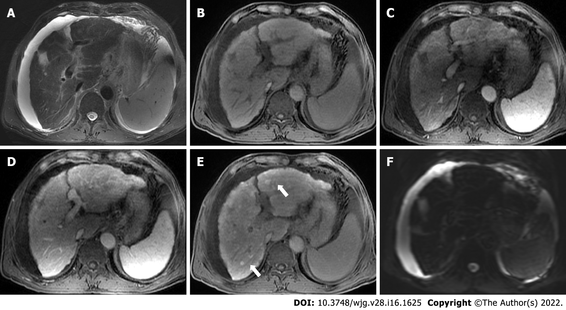
Figure 2 Gadoxetate-enhanced magnetic resonance images of a 70-year-old man with chronic hepatitis B.
T2-weighted image (A) shows signal loss of the liver parenchyma, suggesting iron overload. T1-weighted pre-contrast (B), arterial phase (C), and portal venous phase (D) images show nodular contour and patchy enhancement of the liver parenchyma. Hepatobiliary phase image demonstrates diffuse hyperintense nodules (E, black arrows) without diffusion restriction on diffusion-weighted imaging (F), indicating regenerative nodules. Moderate ascites was also noted.
- Citation: Duan T, Jiang HY, Ling WW, Song B. Noninvasive imaging of hepatic dysfunction: A state-of-the-art review. World J Gastroenterol 2022; 28(16): 1625-1640
- URL: https://www.wjgnet.com/1007-9327/full/v28/i16/1625.htm
- DOI: https://dx.doi.org/10.3748/wjg.v28.i16.1625


