Copyright
©The Author(s) 2022.
World J Gastroenterol. Apr 21, 2022; 28(15): 1548-1562
Published online Apr 21, 2022. doi: 10.3748/wjg.v28.i15.1548
Published online Apr 21, 2022. doi: 10.3748/wjg.v28.i15.1548
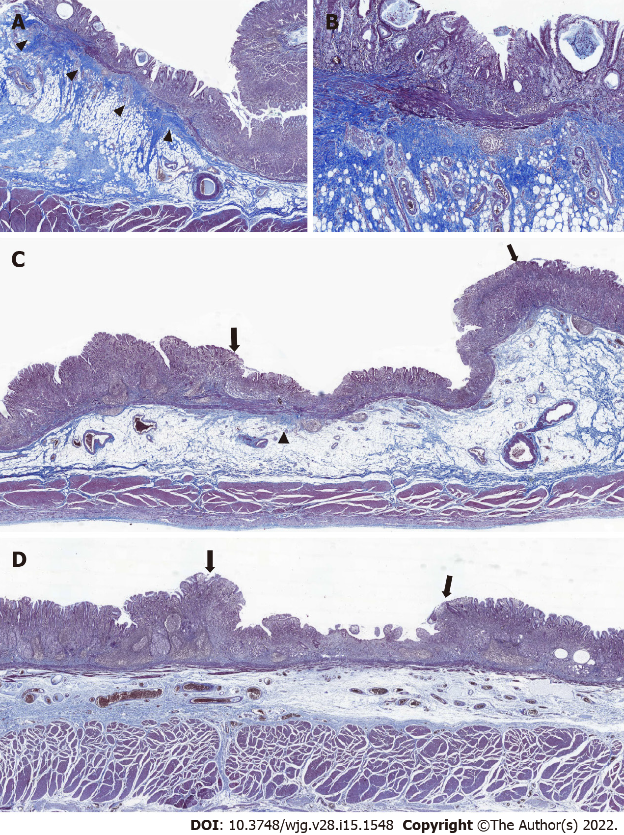
Figure 2 Blurring of muscularis mucosa underneath the tumorous epithelium.
Representative images of tumors with diffuse, focal, and no blurring of muscularis mucosa. A: Diffuse blurring of muscularis mucosa (MM) was prominent enough to localize the tumor at scanning magnification (arrowhead); B: At higher magnification (40´), the thickness of MM appeared irregular due to collagen fibers disrupting the muscle fibers of MM; C: The majority of MM underneath the tumorous epithelium (both ends are marked by arrows) was undisrupted compared with adjacent MM underneath the non-tumorous epithelium, making the foci of MM blurring focal (arrowhead); D: With no blurring of MM, it was difficult to localize the tumor (both ends are marked by arrows) at scanning magnification based on the status of MM.
- Citation: Yoon J, Yoo SY, Park YS, Choi KD, Kim BS, Yoo MW, Lee IS, Yook JH, Kim GH, Na HK, Ahn JY, Lee JH, Jung KW, Kim DH, Song HJ, Lee GH, Jung HY. Reevaluation of the expanded indications in undifferentiated early gastric cancer for endoscopic submucosal dissection. World J Gastroenterol 2022; 28(15): 1548-1562
- URL: https://www.wjgnet.com/1007-9327/full/v28/i15/1548.htm
- DOI: https://dx.doi.org/10.3748/wjg.v28.i15.1548


