Copyright
©The Author(s) 2022.
World J Gastroenterol. Apr 21, 2022; 28(15): 1536-1547
Published online Apr 21, 2022. doi: 10.3748/wjg.v28.i15.1536
Published online Apr 21, 2022. doi: 10.3748/wjg.v28.i15.1536
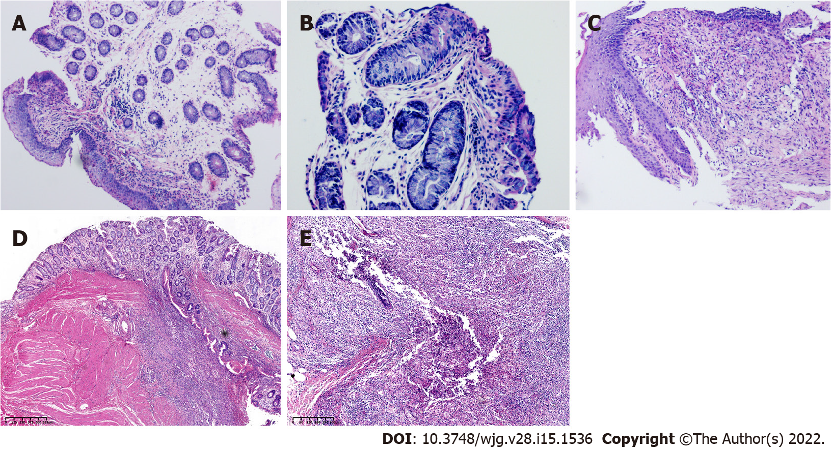
Figure 5 The histological characteristics of a fistula tract.
A: Magnification ×100; B: Magnification ×200; C: Magnification ×100. The early histological changes of fistula are shown; D and E: Longitudinal sections; histological results (rabbits with setons inserted 21 d) showing the inflamed fistula tract. The fistula lumen is visible with internal (digestive side) and external (perineal skin with adipocytes) orifices. There were local inflammatory signs of suppurative inflammation with abscess formation around the fistula.
- Citation: Lu SS, Liu WJ, Niu QY, Huo CY, Cheng YQ, Wang EJ, Li RN, Feng FF, Cheng YM, Liu R, Huang J. Establishing a rabbit model of perianal fistulizing Crohn’s disease. World J Gastroenterol 2022; 28(15): 1536-1547
- URL: https://www.wjgnet.com/1007-9327/full/v28/i15/1536.htm
- DOI: https://dx.doi.org/10.3748/wjg.v28.i15.1536


