Copyright
©The Author(s) 2022.
World J Gastroenterol. Apr 21, 2022; 28(15): 1536-1547
Published online Apr 21, 2022. doi: 10.3748/wjg.v28.i15.1536
Published online Apr 21, 2022. doi: 10.3748/wjg.v28.i15.1536
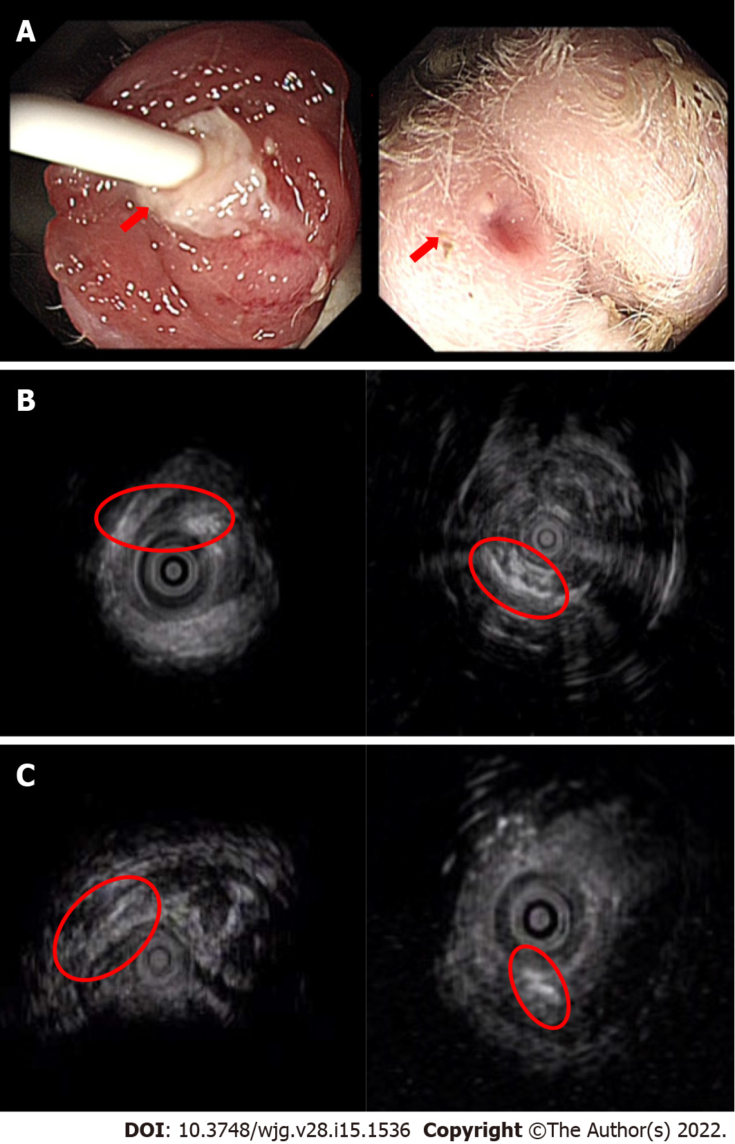
Figure 4 Visualization of a transsphincteric anal fistula via endoscopic ultrasonography.
A: The internal and external openings of the experimental fistula can be directly observed; B: All rabbits with a 21-d insertion time of the surgical thread showed a complete fistula; C: The rabbits with short thread insertion times had different degrees of fistula healing or the disappearance of internal and external fistulas. D0: Day 0; D7: Day 7.
- Citation: Lu SS, Liu WJ, Niu QY, Huo CY, Cheng YQ, Wang EJ, Li RN, Feng FF, Cheng YM, Liu R, Huang J. Establishing a rabbit model of perianal fistulizing Crohn’s disease. World J Gastroenterol 2022; 28(15): 1536-1547
- URL: https://www.wjgnet.com/1007-9327/full/v28/i15/1536.htm
- DOI: https://dx.doi.org/10.3748/wjg.v28.i15.1536


