Copyright
©The Author(s) 2022.
World J Gastroenterol. Apr 21, 2022; 28(15): 1536-1547
Published online Apr 21, 2022. doi: 10.3748/wjg.v28.i15.1536
Published online Apr 21, 2022. doi: 10.3748/wjg.v28.i15.1536
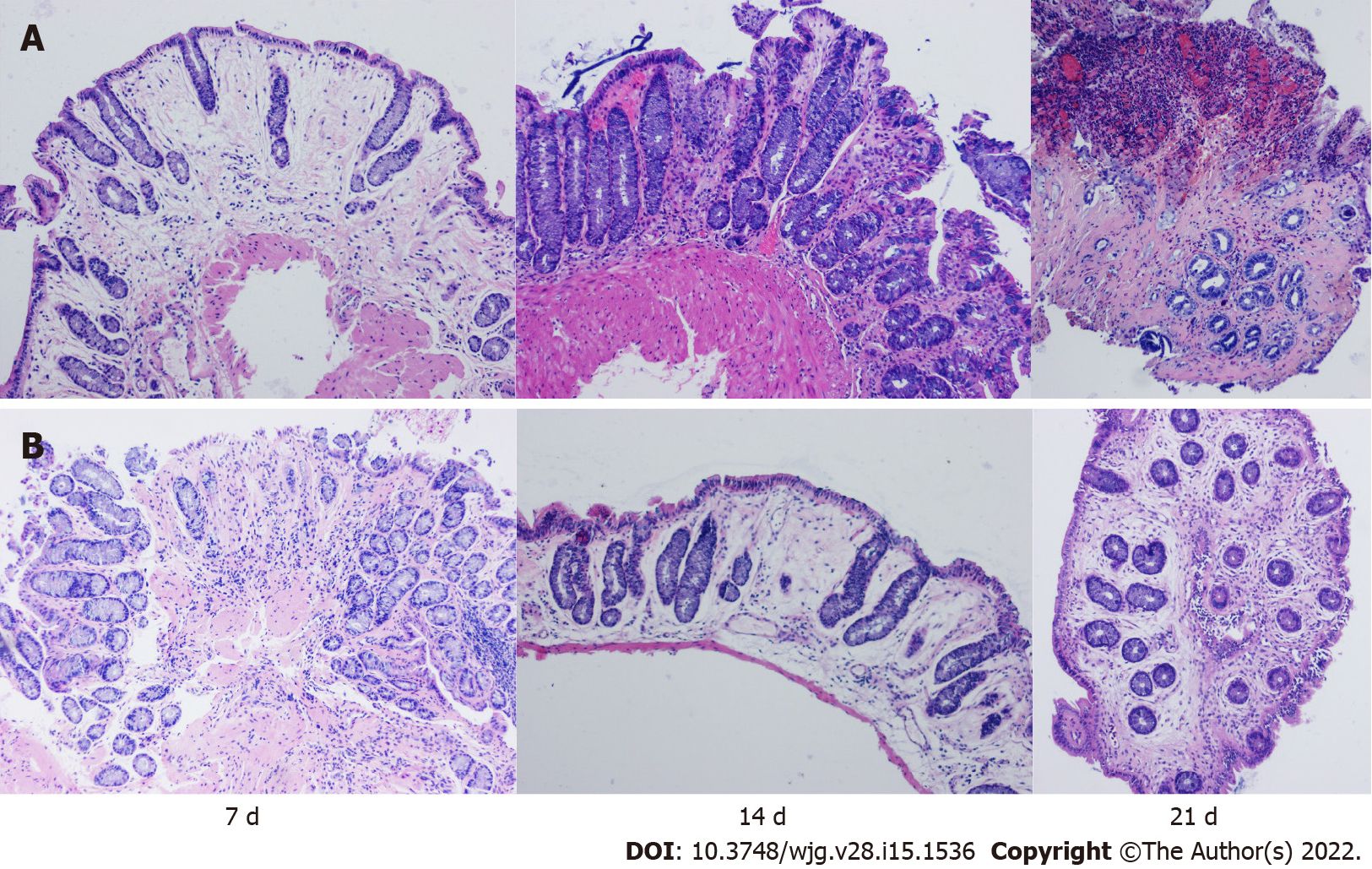
Figure 3 The rabbits in groups A and B underwent three consecutive endoscopic biopsies, and samples were stained with haematoxylin-eosin.
A: Group A exhibited mild to moderate inflammation on day 7, manifested by fewer crypts, marked mucosal oedema, and minimal inflammatory cell infiltration; Group A exhibited fibrosis, hyperaemia, and moderate inflammatory cell infiltration on day 14, and severe inflammatory mucosal ulceration on day 21; B: On the 7th day, there was a higher number of shallow crypts, less oedema and less inflammatory cell infiltration in group B. On the 14th day, there was severe oedema, decreased crypts and less inflammatory cell infiltration in the mucosa, and on the 21st day, there was moderate fibrosis and oedema, mild inflammatory cell infiltration and a complete crypt structure (magnification ×100).
- Citation: Lu SS, Liu WJ, Niu QY, Huo CY, Cheng YQ, Wang EJ, Li RN, Feng FF, Cheng YM, Liu R, Huang J. Establishing a rabbit model of perianal fistulizing Crohn’s disease. World J Gastroenterol 2022; 28(15): 1536-1547
- URL: https://www.wjgnet.com/1007-9327/full/v28/i15/1536.htm
- DOI: https://dx.doi.org/10.3748/wjg.v28.i15.1536


