Copyright
©The Author(s) 2022.
World J Gastroenterol. Apr 21, 2022; 28(15): 1536-1547
Published online Apr 21, 2022. doi: 10.3748/wjg.v28.i15.1536
Published online Apr 21, 2022. doi: 10.3748/wjg.v28.i15.1536
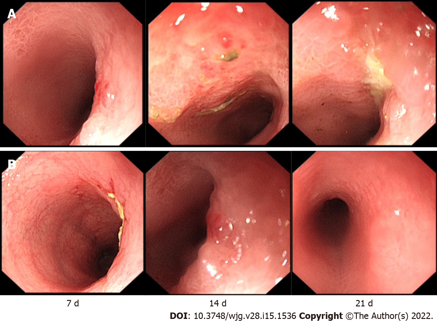
Figure 2 The rabbits in groups A and B underwent endoscopy 3 times.
A: In group A, mild mucosal erosion was observed on the 7th day. On the 14th day, a large area of mucosal oedema and erosion appeared. On the 21st day, the surrounding mucosa was swollen, and a central ulcer had formed; B: In group B, ulceration occurred on the 7th day. On the 14th day, the mucosa was swollen, and the surface was hyperaemic and eroded. On the 21st day, the main manifestation was local congestion.
- Citation: Lu SS, Liu WJ, Niu QY, Huo CY, Cheng YQ, Wang EJ, Li RN, Feng FF, Cheng YM, Liu R, Huang J. Establishing a rabbit model of perianal fistulizing Crohn’s disease. World J Gastroenterol 2022; 28(15): 1536-1547
- URL: https://www.wjgnet.com/1007-9327/full/v28/i15/1536.htm
- DOI: https://dx.doi.org/10.3748/wjg.v28.i15.1536


