Copyright
©The Author(s) 2022.
World J Gastroenterol. Apr 14, 2022; 28(14): 1479-1493
Published online Apr 14, 2022. doi: 10.3748/wjg.v28.i14.1479
Published online Apr 14, 2022. doi: 10.3748/wjg.v28.i14.1479
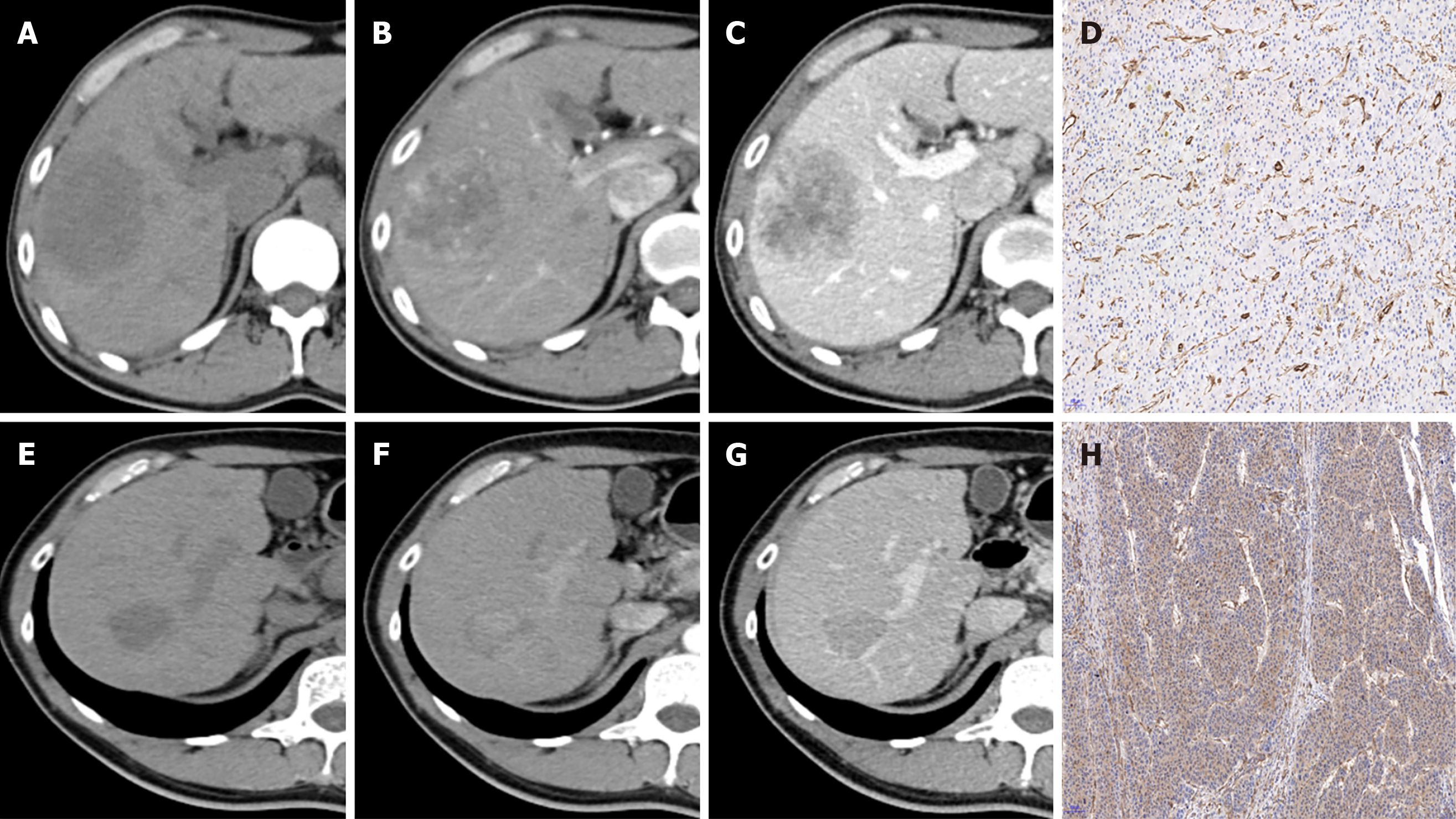
Figure 3 Representative images of contrast-enhanced computed tomography and β-Arrestin1 phosphorylation (magnification, × 100).
A: CT images of a 45-year-old man with a 6.3-cm hepatocellular carcinoma (HCC) in the right liver lobe in the plain phase; B: The tumor shows heterogeneous hyperenhancement in the arterial phase; C: The tumor shows washout at the portal venous phase with intratumor necrosis, an ill-defined capsule and a non-smooth tumor margin. D: Immunohistochemical staining shows a β-arrestin1 phosphorylation-negative status at 100× magnification.
- Citation: Che F, Xu Q, Li Q, Huang ZX, Yang CW, Wang LY, Wei Y, Shi YJ, Song B. Radiomics signature: A potential biomarker for β-arrestin1 phosphorylation prediction in hepatocellular carcinoma. World J Gastroenterol 2022; 28(14): 1479-1493
- URL: https://www.wjgnet.com/1007-9327/full/v28/i14/1479.htm
- DOI: https://dx.doi.org/10.3748/wjg.v28.i14.1479


