Copyright
©The Author(s) 2022.
World J Gastroenterol. Mar 21, 2022; 28(11): 1113-1122
Published online Mar 21, 2022. doi: 10.3748/wjg.v28.i11.1113
Published online Mar 21, 2022. doi: 10.3748/wjg.v28.i11.1113
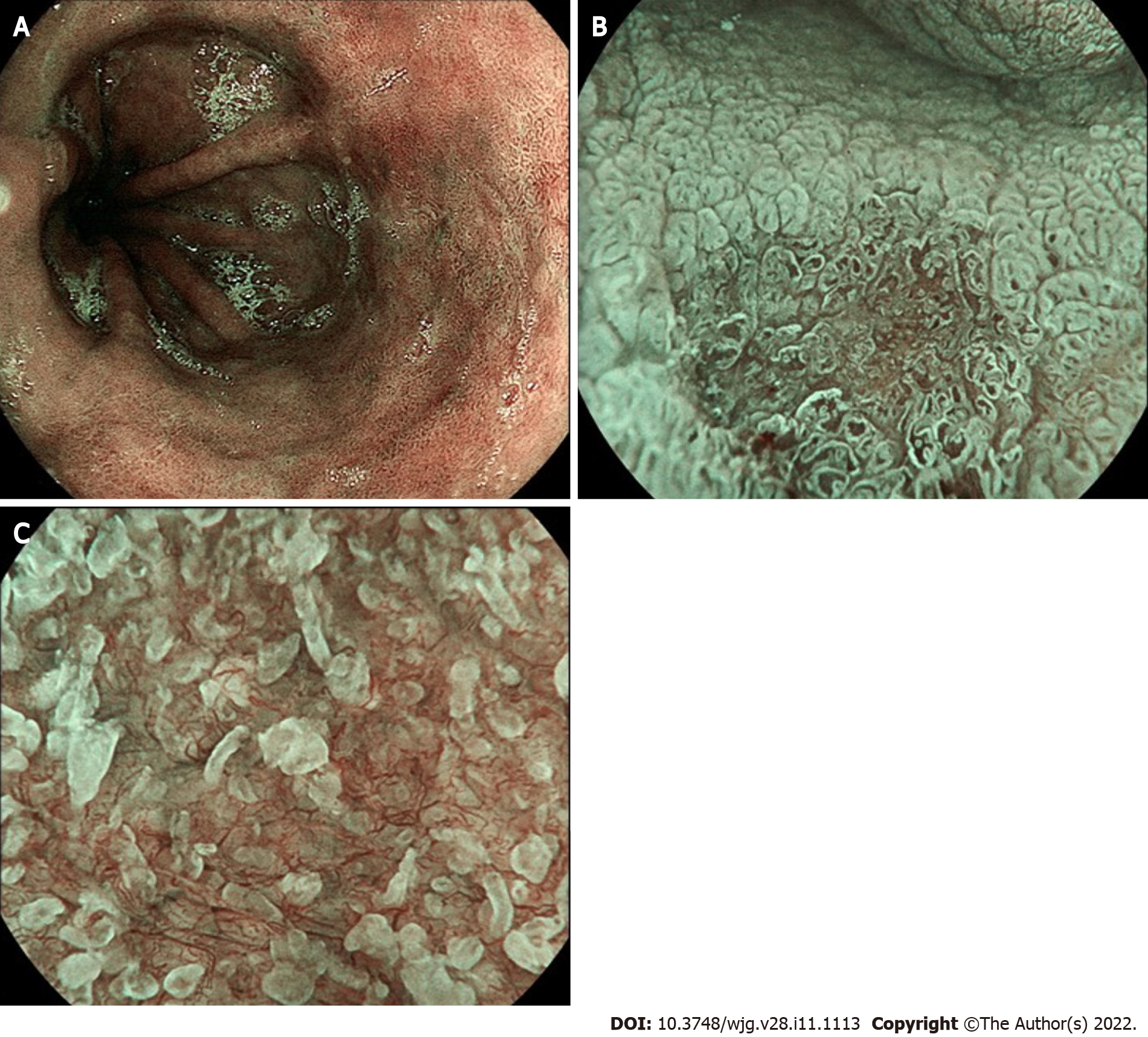
Figure 4 Blue light imaging appearance of Barrett’s mucosa.
A: Non-dysplastic mucosa; B: Irregular vascular pattern of a slightly depressed (Paris 0-IIc) high-grade dysplastic lesion; C: Detail of irregular mucosal and vascular pattern after zoom magnification.
- Citation: Spadaccini M, Vespa E, Chandrasekar VT, Desai M, Patel HK, Maselli R, Fugazza A, Carrara S, Anderloni A, Franchellucci G, De Marco A, Hassan C, Bhandari P, Sharma P, Repici A. Advanced imaging and artificial intelligence for Barrett's esophagus: What we should and soon will do . World J Gastroenterol 2022; 28(11): 1113-1122
- URL: https://www.wjgnet.com/1007-9327/full/v28/i11/1113.htm
- DOI: https://dx.doi.org/10.3748/wjg.v28.i11.1113


