Copyright
©The Author(s) 2022.
World J Gastroenterol. Mar 14, 2022; 28(10): 976-984
Published online Mar 14, 2022. doi: 10.3748/wjg.v28.i10.976
Published online Mar 14, 2022. doi: 10.3748/wjg.v28.i10.976
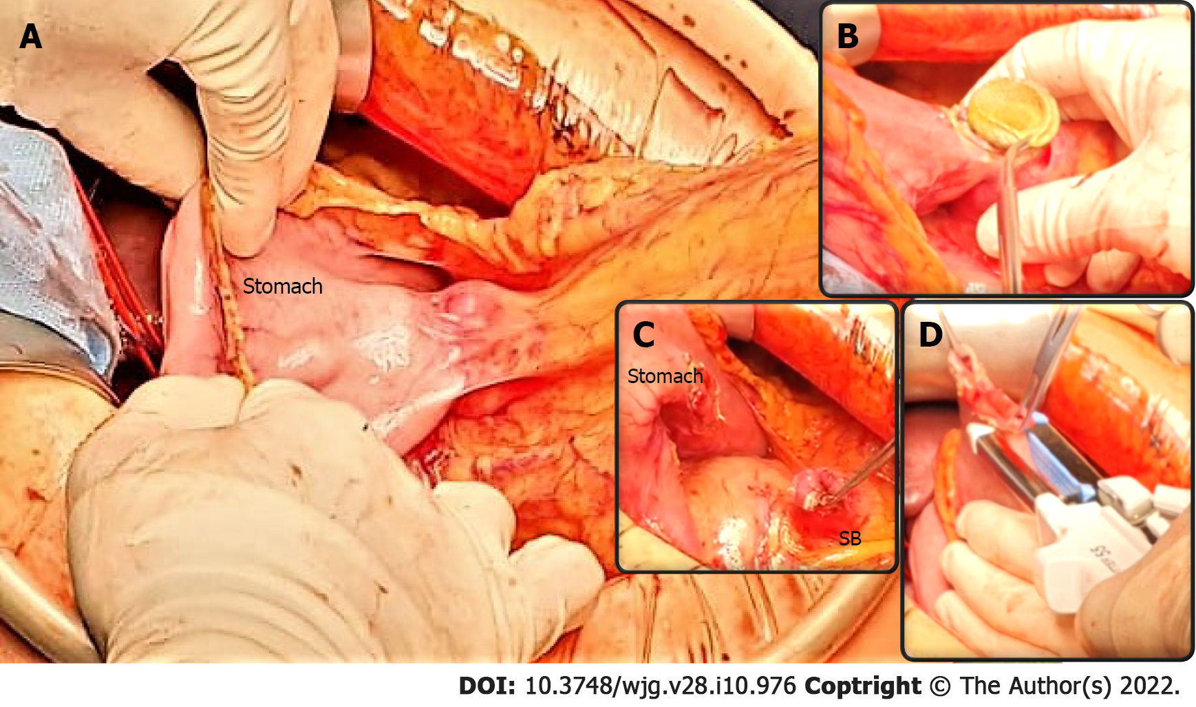
Figure 4 Pancreaticoduodenectomy following endoscopic ultrasound-guided double bypass.
A: The endoscopic ultrasound-guided gastrojejunostomy site is easily identified and a stable gastrojejunal anastomosis is visible (underlined by blue curves) between the stomach and the small bowel (SB); B-C: The endoscopic ultrasound-guided gastrojejunostomy site is opened with diathermic coagulation, the lumen apposing metal stent is removed (B), and the anastomosis cut (C); D: The SB is prepared for gastroenteric anastomosis, while the gastric defect will be closed using staplers. A classic pylorus-preserving pancreaticoduodenectomy is achievable.
- Citation: Vanella G, Tamburrino D, Capurso G, Bronswijk M, Reni M, Dell'Anna G, Crippa S, Van der Merwe S, Falconi M, Arcidiacono PG. Feasibility of therapeutic endoscopic ultrasound in the bridge-to-surgery scenario: The example of pancreatic adenocarcinoma. World J Gastroenterol 2022; 28(10): 976-984
- URL: https://www.wjgnet.com/1007-9327/full/v28/i10/976.htm
- DOI: https://dx.doi.org/10.3748/wjg.v28.i10.976


