Copyright
©The Author(s) 2022.
World J Gastroenterol. Mar 14, 2022; 28(10): 1078-1084
Published online Mar 14, 2022. doi: 10.3748/wjg.v28.i10.1078
Published online Mar 14, 2022. doi: 10.3748/wjg.v28.i10.1078
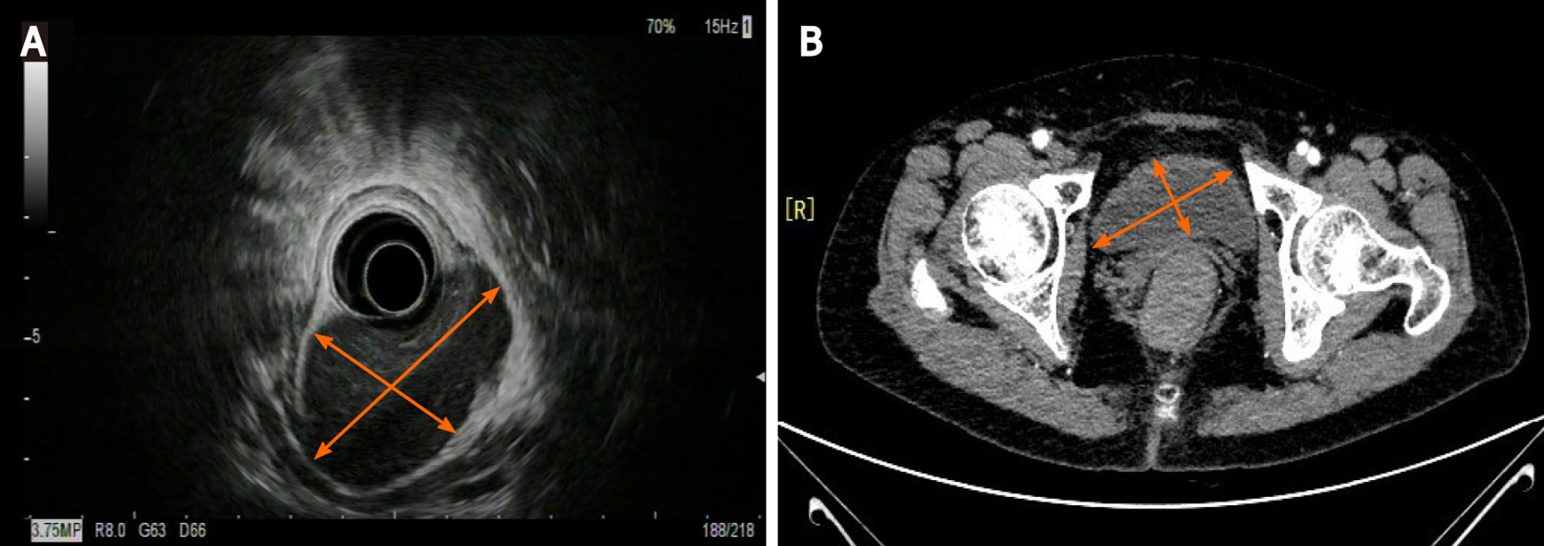
Figure 2 Endoscopic ultrasonography (EUS) and computed tomography (CT) of the lesion.
A: EUS showing a 50 mm, low echo, submucosal lesion in the low rectum; B: Abdominal enhanced CT imaging revealing a 50 mm × 30 mm soft tissue mass with defined boundary, uniform density and moderate enhancement.
- Citation: Li FY, Zhang XL, Zhang QD, Wang YH. Successful treatment of an enormous rectal mucosa-associated lymphoid tissue lymphoma by endoscopic full-thickness resection: A case report. World J Gastroenterol 2022; 28(10): 1078-1084
- URL: https://www.wjgnet.com/1007-9327/full/v28/i10/1078.htm
- DOI: https://dx.doi.org/10.3748/wjg.v28.i10.1078


