Copyright
©The Author(s) 2022.
World J Gastroenterol. Mar 14, 2022; 28(10): 1055-1066
Published online Mar 14, 2022. doi: 10.3748/wjg.v28.i10.1055
Published online Mar 14, 2022. doi: 10.3748/wjg.v28.i10.1055
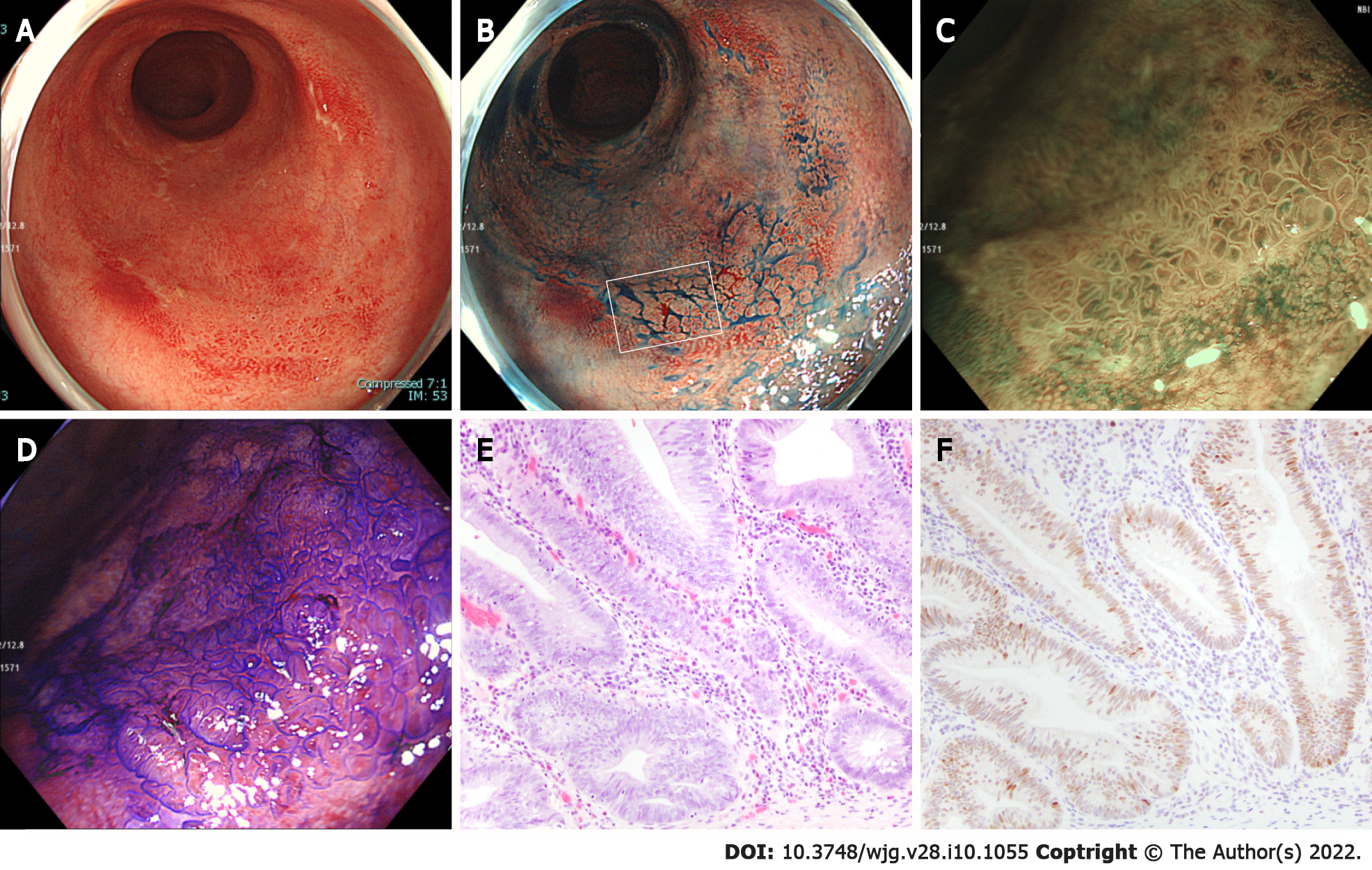
Figure 1 Endoscopic features of ulcerative colitis-associated neoplasms misdiagnosed by all endoscopists.
A: White-light imaging reveals a flat elevated lesion in the rectum; B: Chromoendoscopy with indigo carmine shows a clear lesion border; C: Magnifying endoscopy with narrow-band imaging of box in (B) shows regular surface and vascular patterns, which were classified by all endoscopists as Japan Narrow-Band Imaging Expert Team classification type 2A; D: Magnifying endoscopy with crystal violet chromoendoscopy of box in (B) reveals relatively uniform villous structures, which were classified by all endoscopists as pit pattern type IV; E: Pathological examination of the resected specimen by endoscopic submucosal dissection shows architectural atypia. This lesion was pathologically diagnosed as high-grade dysplasia (hematoxylin and eosin staining, original magnification × 50); F: Immunohistochemistry for p53 on serial section of (E).
- Citation: Kida Y, Yamamura T, Maeda K, Sawada T, Ishikawa E, Mizutani Y, Kakushima N, Furukawa K, Ishikawa T, Ohno E, Kawashima H, Nakamura M, Ishigami M, Fujishiro M. Diagnostic performance of endoscopic classifications for neoplastic lesions in patients with ulcerative colitis: A retrospective case-control study. World J Gastroenterol 2022; 28(10): 1055-1066
- URL: https://www.wjgnet.com/1007-9327/full/v28/i10/1055.htm
- DOI: https://dx.doi.org/10.3748/wjg.v28.i10.1055


