Copyright
©The Author(s) 2021.
World J Gastroenterol. Feb 28, 2021; 27(8): 751-759
Published online Feb 28, 2021. doi: 10.3748/wjg.v27.i8.751
Published online Feb 28, 2021. doi: 10.3748/wjg.v27.i8.751
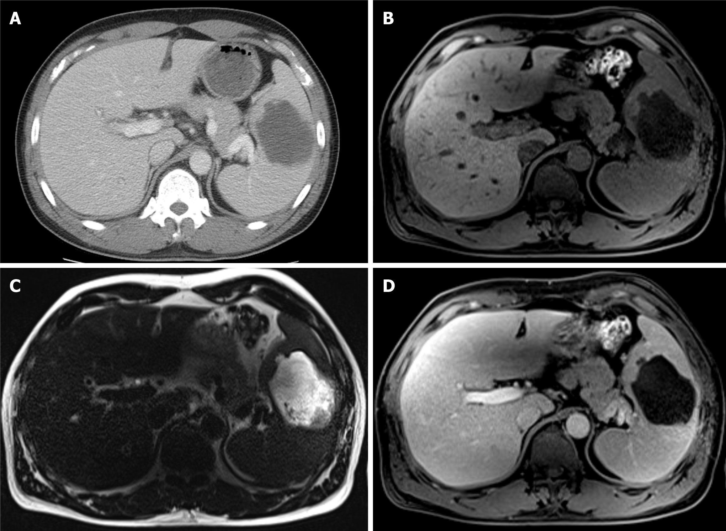
Figure 3 Abdominal computed tomography and magnetic resonance imaging at the readmission.
A: Abdominal computed tomography revealed a 7 cm-sized low-density lesion; B: T1-weighted magnetic resonance imaging (MRI) revealed a 7cm-sized hypointense mass with a lower signal compared to the initial MRI; C: T2-weighted MRI revealed much higher signal intensity of this lesion compared to the initial MRI with non-enhancing debris or necrotic portions inside; D: Gadolinium-enhanced T1-weighted image revealed a much lower signal lesion with minimal peripheral enhancement suspecting a capsule development, suggestive for abscess formation.
- Citation: Cho SY, Cho E, Park CH, Kim HJ, Koo JY. Septic shock due to Granulicatella adiacens after endoscopic ultrasound-guided biopsy of a splenic mass: A case report. World J Gastroenterol 2021; 27(8): 751-759
- URL: https://www.wjgnet.com/1007-9327/full/v27/i8/751.htm
- DOI: https://dx.doi.org/10.3748/wjg.v27.i8.751


