Copyright
©The Author(s) 2021.
World J Gastroenterol. Feb 28, 2021; 27(8): 751-759
Published online Feb 28, 2021. doi: 10.3748/wjg.v27.i8.751
Published online Feb 28, 2021. doi: 10.3748/wjg.v27.i8.751
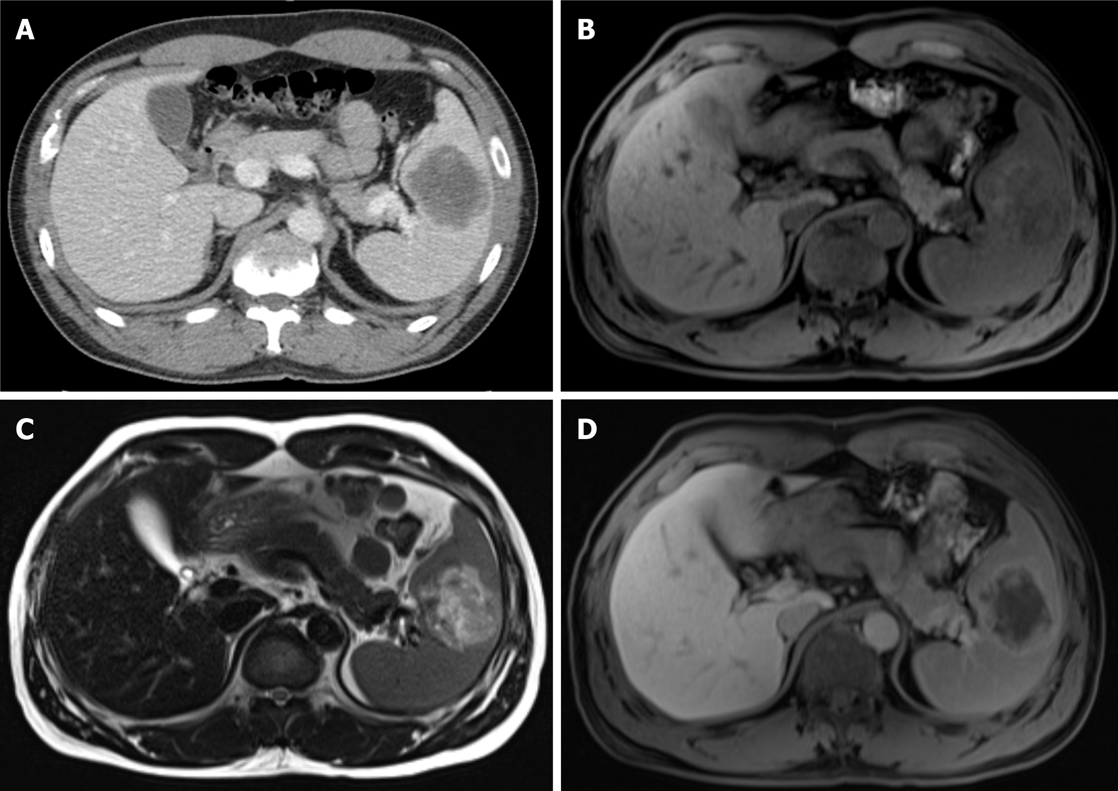
Figure 1 Initial abdominal computed tomography and magnetic resonance imaging findings.
A: Post-contrast phases of abdominal computed tomography showed a 6 cm-sized hypodense mass with peripheral enhancing rim in the spleen; B: T1-weighted abdominal magnetic resonance imaging showed a 6 cm-sized heterogeneously hypointense splenic mass; C: T2-weighted image showed heterogeneous hyperintense signal of this lesion; D: Gadolinium-enhanced T1-weighted image showed peripheral enhancement and a central hypo-enhancing lesion.
- Citation: Cho SY, Cho E, Park CH, Kim HJ, Koo JY. Septic shock due to Granulicatella adiacens after endoscopic ultrasound-guided biopsy of a splenic mass: A case report. World J Gastroenterol 2021; 27(8): 751-759
- URL: https://www.wjgnet.com/1007-9327/full/v27/i8/751.htm
- DOI: https://dx.doi.org/10.3748/wjg.v27.i8.751


