Copyright
©The Author(s) 2021.
World J Gastroenterol. Dec 28, 2021; 27(48): 8227-8241
Published online Dec 28, 2021. doi: 10.3748/wjg.v27.i48.8227
Published online Dec 28, 2021. doi: 10.3748/wjg.v27.i48.8227
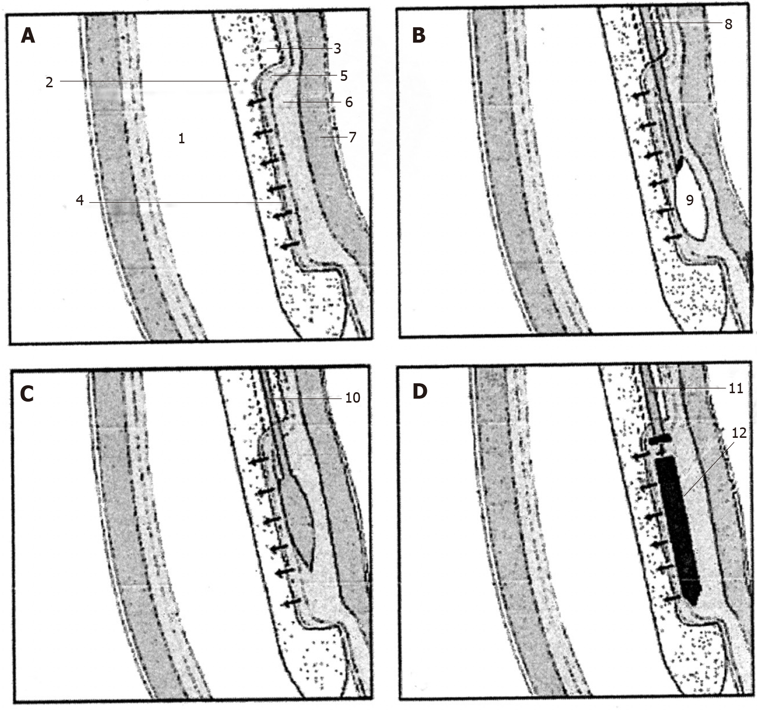
Figure 3 Extremity of the special endoesophageal probe positioned at the LES level in a sequence of operations for the deployment of a magnetic plaque seen in profile.
A: The mucosa of the distal esophagus is sucked onto the perforated wall of the operative chamber; B: The needle injects milliliters of saline solution to create a blister in the submucosa; C: The end of the catheter with a blunted bolt creates a pouch in the submucosa; D: The magnetic plaque (seen in profile) is pushed into the pouch. 1: Esophageal lumen; 2: Delivery probe; 3: Deployment channel; 4: Perforated wall of the aspiration chamber; 5: Mucosal layer; 6: Submucosal layer; 7: Muscular layer; 8: Needle-catheter; 9: Saline solution; 10: Bolt-catheter; 11: Magnetic plaque seen in profile. A-D: Citation: Bortolotti M, Grandis A, Mazzero G. A novel endoesophageal magnetic device to prevent gastroesophageal reflux. Surg Endosc 2009; 4: 885-9. Copyright© The Authors 2020. Published by Springer Nature. The authors obtained permission for use of the figure from Springer Nature (Supplementary material).
- Citation: Bortolotti M. Magnetic challenge against gastroesophageal reflux . World J Gastroenterol 2021; 27(48): 8227-8241
- URL: https://www.wjgnet.com/1007-9327/full/v27/i48/8227.htm
- DOI: https://dx.doi.org/10.3748/wjg.v27.i48.8227


