Copyright
©The Author(s) 2021.
World J Gastroenterol. Dec 14, 2021; 27(46): 7866-7893
Published online Dec 14, 2021. doi: 10.3748/wjg.v27.i46.7866
Published online Dec 14, 2021. doi: 10.3748/wjg.v27.i46.7866
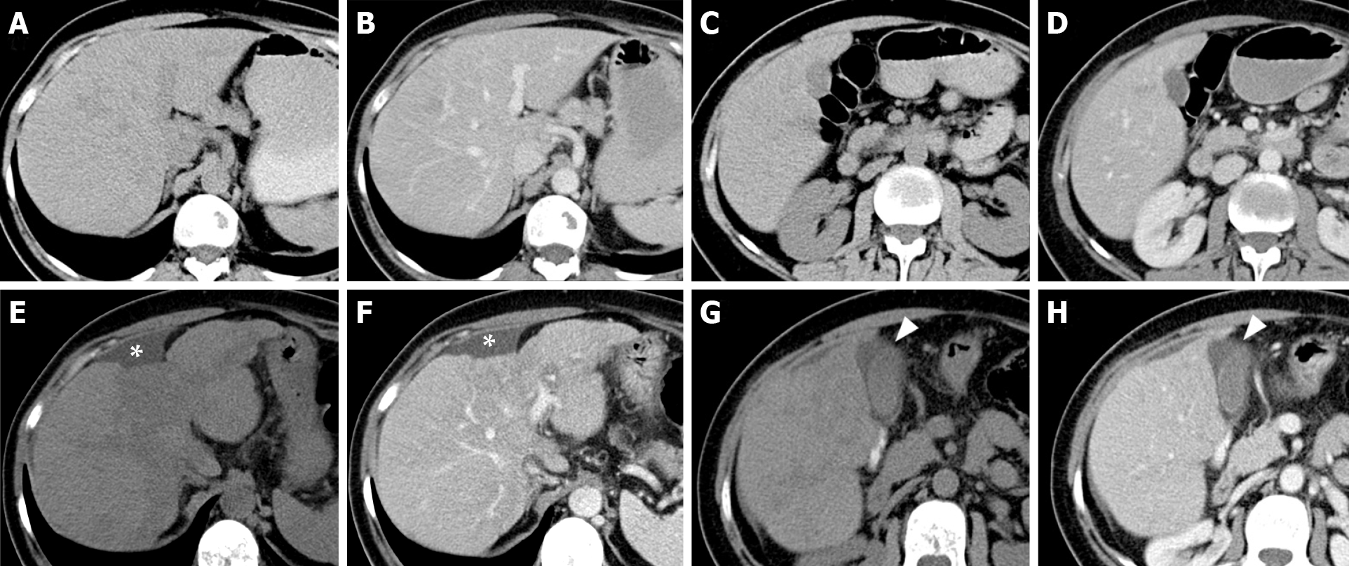
Figure 14 Early features of pseudocirrhosis in patients with metastatic breast cancer treated with gemcitabine for 12 mo.
A-D: On axial unenhanced (A, C) and contrast-enhanced portal (B, D) computed tomography (CT) scan images, executed prior chemotherapy, the liver presents a regular volume, morphology and a smooth surface. No signs of ascites are present; E-H: On CT exam after chemotherapy (12 mo) at the same levels, in the same phases, fatty changes of the liver parenchyma, reduction of the hepatic volume with relative hypertrophy of the left lobe, irregular margins and capsular retraction corresponding to the IV segment (asterisk) were detectable. Peri-hepatic and pericholecystic effusion occurred (arrowhead).
- Citation: Calistri L, Rastrelli V, Nardi C, Maraghelli D, Vidali S, Pietragalla M, Colagrande S. Imaging of the chemotherapy-induced hepatic damage: Yellow liver, blue liver, and pseudocirrhosis. World J Gastroenterol 2021; 27(46): 7866-7893
- URL: https://www.wjgnet.com/1007-9327/full/v27/i46/7866.htm
- DOI: https://dx.doi.org/10.3748/wjg.v27.i46.7866


