Copyright
©The Author(s) 2021.
World J Gastroenterol. Dec 14, 2021; 27(46): 7866-7893
Published online Dec 14, 2021. doi: 10.3748/wjg.v27.i46.7866
Published online Dec 14, 2021. doi: 10.3748/wjg.v27.i46.7866
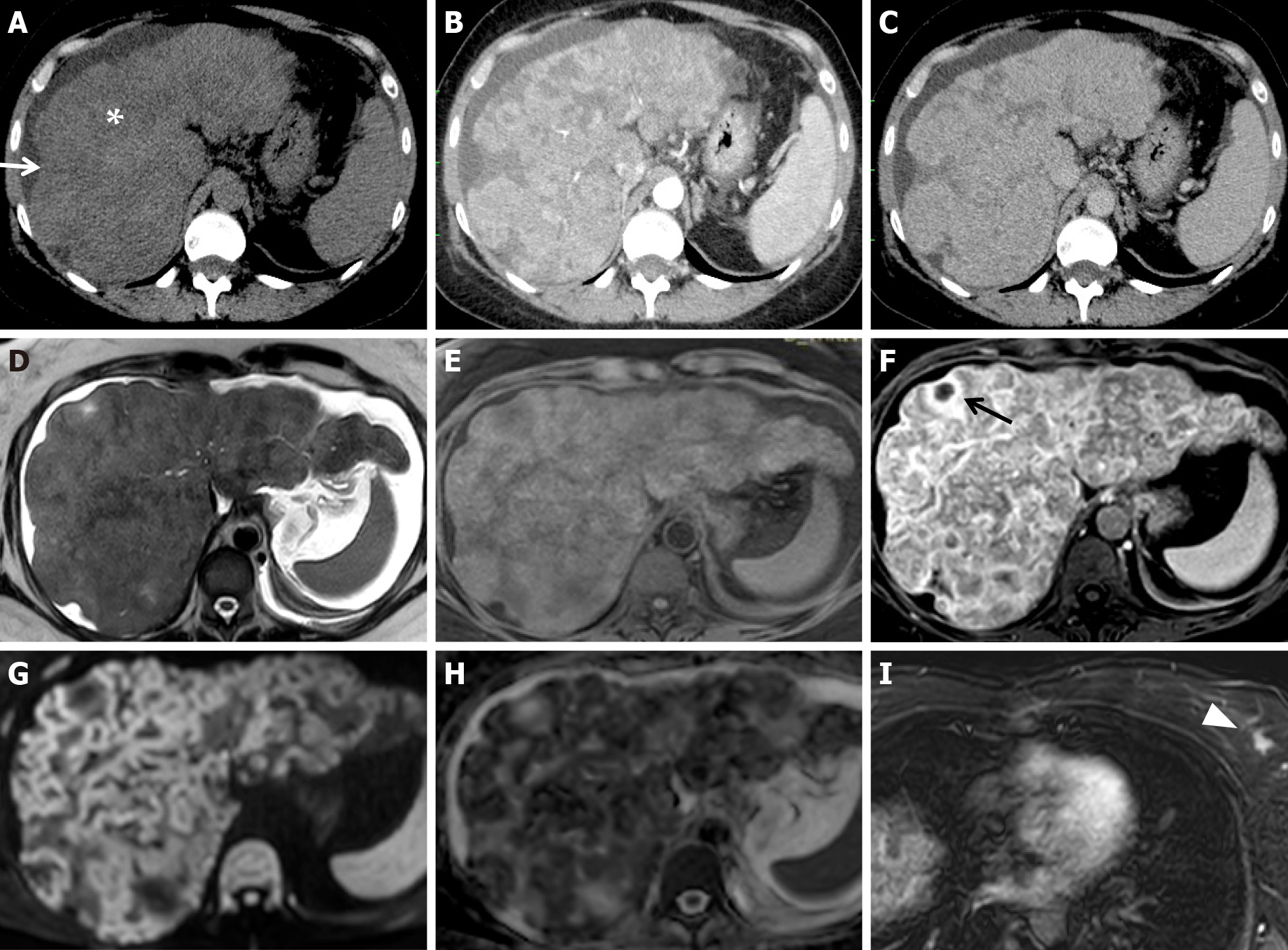
Figure 13 Pathologically proven pseudocirrhosis due to a small breast cancer in a chemotherapy “naïve patient”, having received no chemotherapy.
A-C: On unenhanced (A) computed tomography (CT) axial scans, a lobulated liver contour with retraction of the capsular surface (white arrow), low-attenuation parenchymal areas, and ascites (white asterisk) are seen. On arterial (B) and portal (C) CT axial scans, architectural disorder and heterogeneous contrast enhancement are detectable; D-I: On magnetic resonance, the presence of ascites is confirmed on T2w images (D). Profound structural and architectural changes due to the presence of coarse nodules separated by areas of fibrosis in an unenhanced fat sat gradient echo 3D T1w image (E) and a contrast-enhanced phase T1w image at equilibrium (F) are visible; various confluent nodules with irregular hyperintense rims on high b-value diffusion-weighted images (G) and low signal intensity in apparent diffusion coefficient map value (H) were observed. A small necrotic area inside a nodule is indicated in F (black arrow). One small left breast cancer nodule (white arrowhead) on a contrast-enhanced T1w image is visible in the arterial phase (I).
- Citation: Calistri L, Rastrelli V, Nardi C, Maraghelli D, Vidali S, Pietragalla M, Colagrande S. Imaging of the chemotherapy-induced hepatic damage: Yellow liver, blue liver, and pseudocirrhosis. World J Gastroenterol 2021; 27(46): 7866-7893
- URL: https://www.wjgnet.com/1007-9327/full/v27/i46/7866.htm
- DOI: https://dx.doi.org/10.3748/wjg.v27.i46.7866


