Copyright
©The Author(s) 2021.
World J Gastroenterol. Dec 14, 2021; 27(46): 7866-7893
Published online Dec 14, 2021. doi: 10.3748/wjg.v27.i46.7866
Published online Dec 14, 2021. doi: 10.3748/wjg.v27.i46.7866
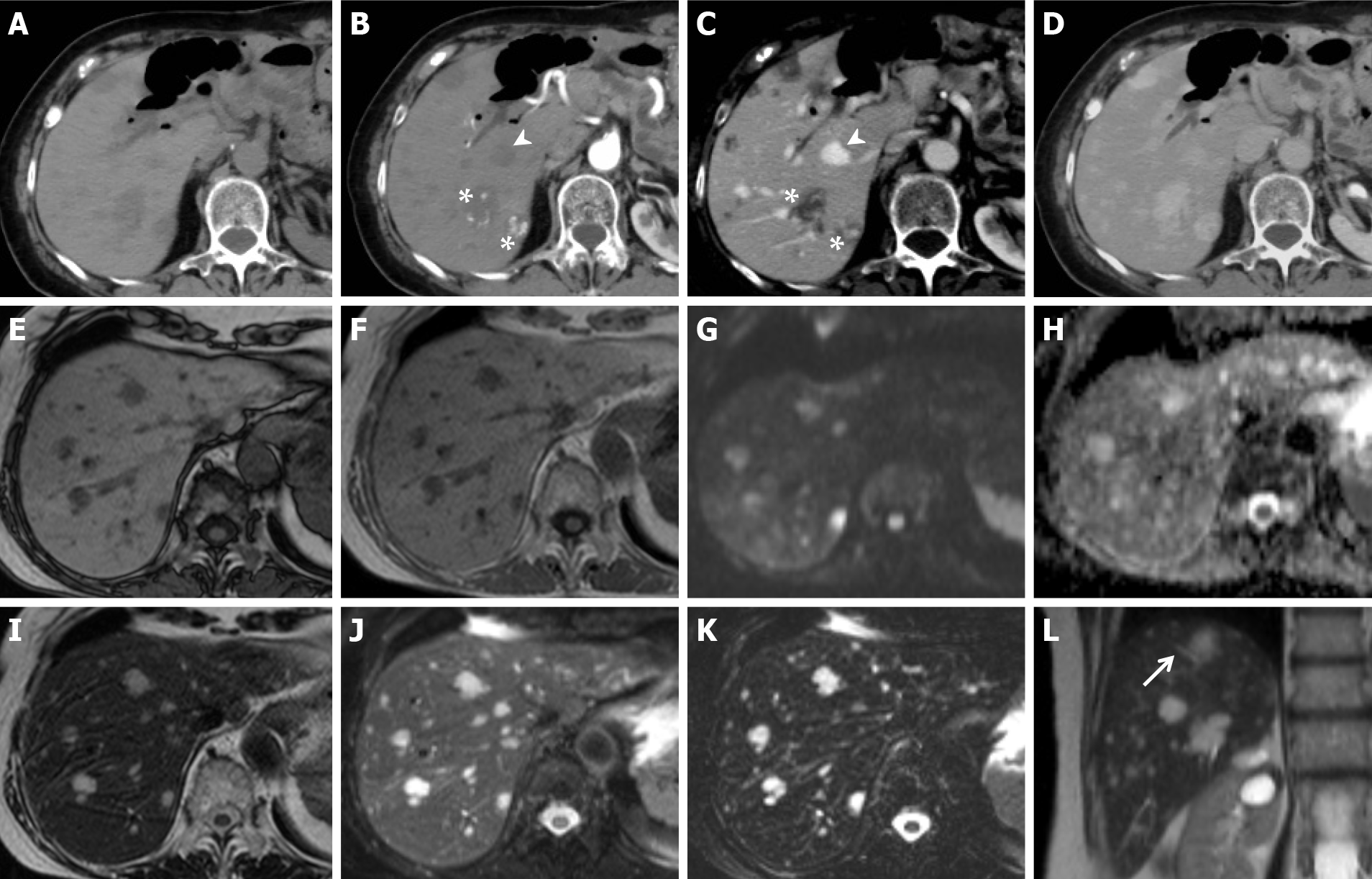
Figure 10 Secondary idiopathic multiple peliotic lesions in patients with a history of 6-mercaptopurine treatment for leukemia.
A-D: Contrast-enhanced computed tomography shows multiple lesions, hypodense on unenhanced scan (A) with dystrophic calcifications and hyperdense foci, probably secondary to hemorrhage. On dynamic imaging (B, axial arterial phase; C, axial portal phase), the lesions present centripetal (arrowhead) or centrifugal (asterisk) globular contrast enhancement without signs of washout. In the delayed phase (D), they appear isodense compared with the hepatic parenchyma; E-L: Magnetic resonance confirming the presence of hypointense lesions on T1w images (E-F) and hyperintense lesions on T2w images (I, J and L, arrow), which maintain high signal in long echoes echo time 320 ms (K). No signs of altered diffusion (G-H) or mass effects are shown. These characteristics were consistent with multiple peliotic lesions.
- Citation: Calistri L, Rastrelli V, Nardi C, Maraghelli D, Vidali S, Pietragalla M, Colagrande S. Imaging of the chemotherapy-induced hepatic damage: Yellow liver, blue liver, and pseudocirrhosis. World J Gastroenterol 2021; 27(46): 7866-7893
- URL: https://www.wjgnet.com/1007-9327/full/v27/i46/7866.htm
- DOI: https://dx.doi.org/10.3748/wjg.v27.i46.7866


