Copyright
©The Author(s) 2021.
World J Gastroenterol. Dec 14, 2021; 27(46): 7866-7893
Published online Dec 14, 2021. doi: 10.3748/wjg.v27.i46.7866
Published online Dec 14, 2021. doi: 10.3748/wjg.v27.i46.7866
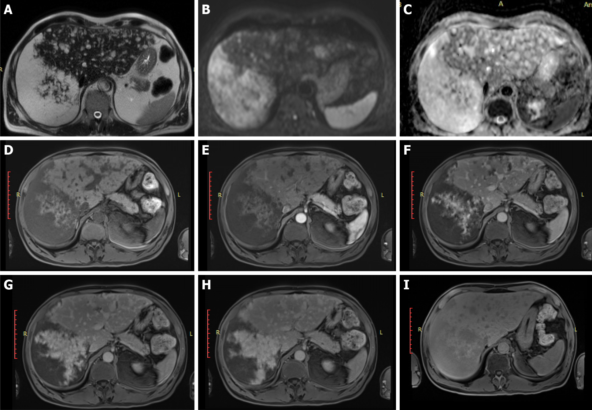
Figure 8 Primary idiopathic diffuse peliosis in patients without a cancer history.
A-C: On magnetic resonance T2w images (A) and on diffusion-weighted imaging (B: High b value; C: Apparent diffusion coefficient map), numerous hemangioma-like lesions are visible, the largest in segments VII-VIII; D-I: After interstitial contrast agent administration, progressive centrifugal enhancement of the lesions was observed (D: Fat sat gradient echo 3D T1w unenhanced image; E, F, G: Arterial, portal and equilibrium phases; H-I: 5 and 30 min after contrast agent administration).
- Citation: Calistri L, Rastrelli V, Nardi C, Maraghelli D, Vidali S, Pietragalla M, Colagrande S. Imaging of the chemotherapy-induced hepatic damage: Yellow liver, blue liver, and pseudocirrhosis. World J Gastroenterol 2021; 27(46): 7866-7893
- URL: https://www.wjgnet.com/1007-9327/full/v27/i46/7866.htm
- DOI: https://dx.doi.org/10.3748/wjg.v27.i46.7866


