Copyright
©The Author(s) 2021.
World J Gastroenterol. Dec 14, 2021; 27(46): 7866-7893
Published online Dec 14, 2021. doi: 10.3748/wjg.v27.i46.7866
Published online Dec 14, 2021. doi: 10.3748/wjg.v27.i46.7866
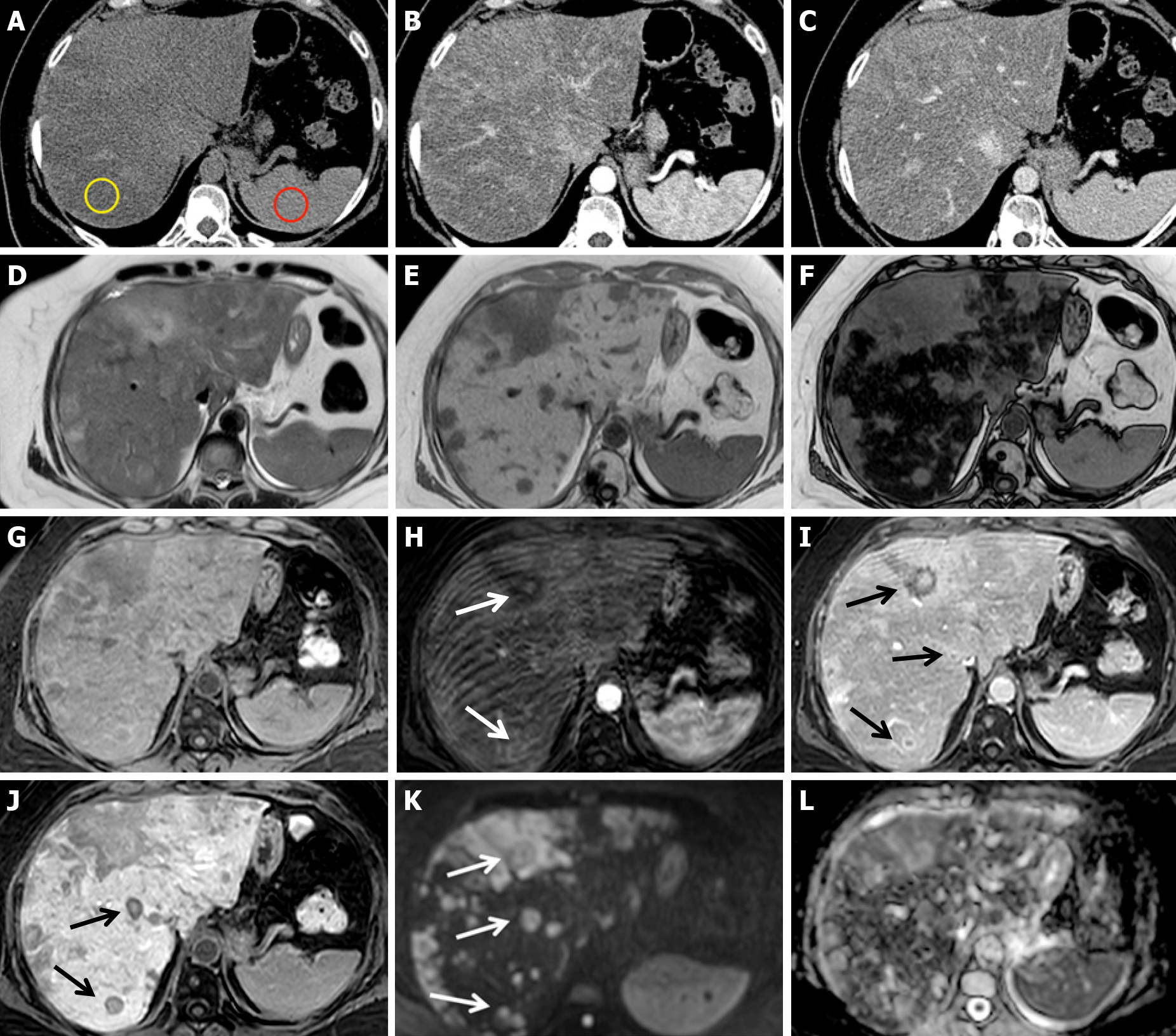
Figure 4 Chemotherapy-associated diffuse steatosis in patients with liver metastatic colorectal cancer 6 mo after the beginning of chemotherapy with 5-fluorouracil + folinic acid + irinotecan.
A-C: Unenhanced computed tomography axial scan (A), arterial (B) and portal phase (C) show severe inhomogeneous hepatic steatosis. Liver attenuation in steatotic areas is 8 HU (yellow ROI), less than that of the spleen (43 HU, red ROI), and the hepatic/splenic attenuation ratio is << 1. No nodules are visible; D-L: On magnetic resonance performed during the following week, gradient echo (GE) T1w in-phase (E) and out-of-phase (F) images confirm severe hepatic steatosis. On unenhanced fat sat GE 3D T1w images (G), arterial (H), portal (I), hepatobiliary (J) phases and high b-value diffusion-weighted images (K), multiple metastases are evident, some of which are characterized by rim enhancement (arrows). The T2-weighted image (D) and apparent diffusion coefficient map (L) are also shown.
- Citation: Calistri L, Rastrelli V, Nardi C, Maraghelli D, Vidali S, Pietragalla M, Colagrande S. Imaging of the chemotherapy-induced hepatic damage: Yellow liver, blue liver, and pseudocirrhosis. World J Gastroenterol 2021; 27(46): 7866-7893
- URL: https://www.wjgnet.com/1007-9327/full/v27/i46/7866.htm
- DOI: https://dx.doi.org/10.3748/wjg.v27.i46.7866


