Copyright
©The Author(s) 2021.
World J Gastroenterol. Nov 21, 2021; 27(43): 7509-7529
Published online Nov 21, 2021. doi: 10.3748/wjg.v27.i43.7509
Published online Nov 21, 2021. doi: 10.3748/wjg.v27.i43.7509
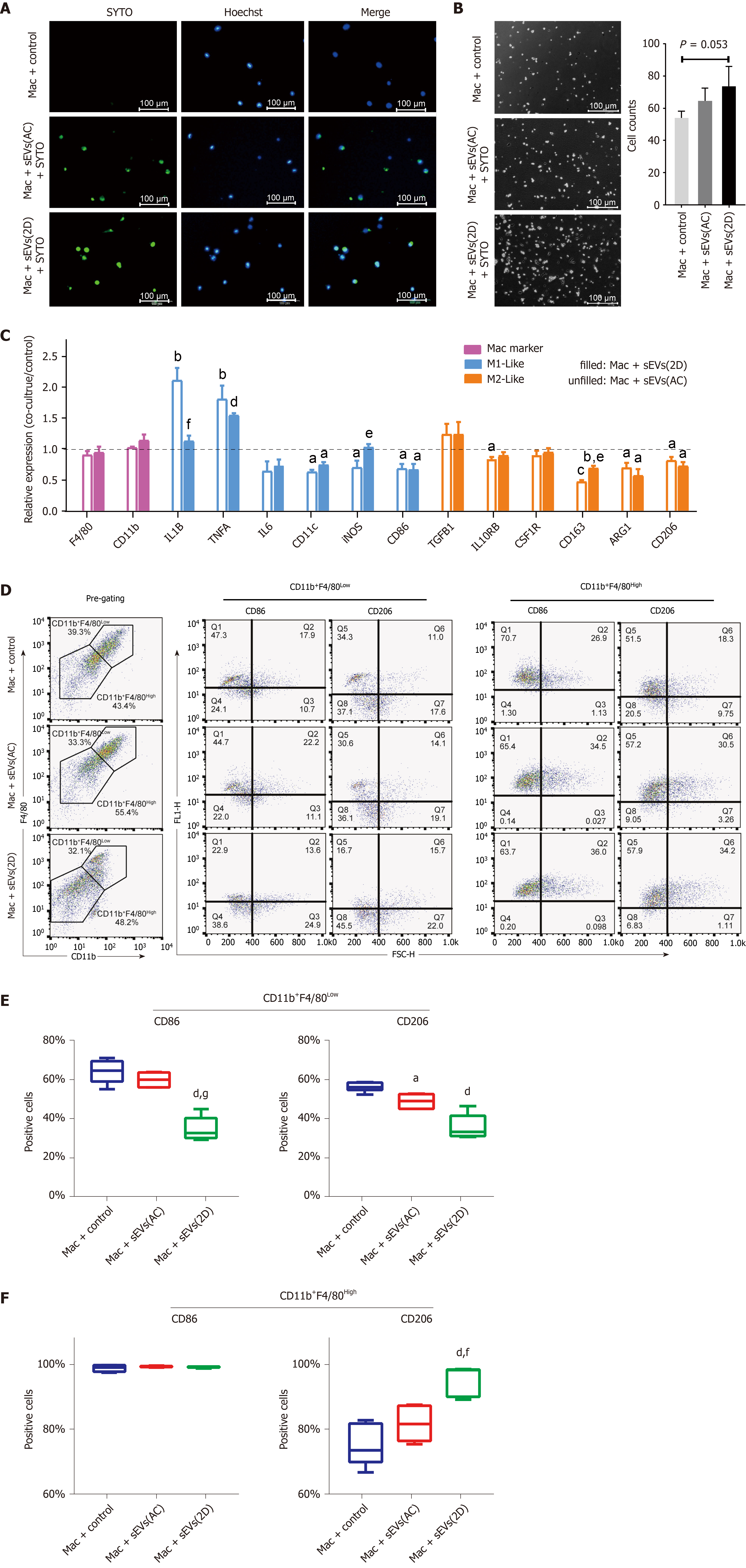
Figure 7 Uptake of serum small extracellular vesicles by hepatic macrophages and subsequent reprogramming.
A: Uptake of SYTO-labeled serum small extracellular vesicles (sEVs) from normal (AC) or acute liver injury (ALI) (2D) mice by primary hepatic macrophages; B: Hepatic macrophages were incubated with AC or 2D serum sEVs for 24 h. The number of attached cells per 200 × field is shown; C: Expression of M1- and M2-like cell surface markers and cytokines in hepatic macrophages incubated with mice serum sEVs. The unfilled column represents macrophages incubated with AC sEVs, and the filled column represents macrophages incubated with 2D sEVs. Compared with the untreated control group, aP < 0.05, bP < 0.01, cP < 0.001, dP < 0.0001; compared with the AC sEV treatment group, eP < 0.05, fP < 0.01; D: Macrophages were defined as CD11b+F4/80Low and CD11b+F4/80High subgroups. The representative images show the percentage of CD86-and CD206-positive cells in each subgroup subjected to the control, AC sEV and 2D sEV treatments; E: CD86- and CD206-positive cells in each subgroup. Compared with the control group, aP < 0.05, dP < 0.0001; compared with the AC sEV treatment group, fP < 0.01, gP < 0.0001. Scale bar = 100 μm. Mac: Macrophage; D: Day; AC: Acute liver injury control; sEVs: Small extracellular vesicles.
- Citation: Lv XF, Zhang AQ, Liu WQ, Zhao M, Li J, He L, Cheng L, Sun YF, Qin G, Lu P, Ji YH, Ji JL. Liver injury changes the biological characters of serum small extracellular vesicles and reprograms hepatic macrophages in mice. World J Gastroenterol 2021; 27(43): 7509-7529
- URL: https://www.wjgnet.com/1007-9327/full/v27/i43/7509.htm
- DOI: https://dx.doi.org/10.3748/wjg.v27.i43.7509


