Copyright
©The Author(s) 2021.
World J Gastroenterol. Nov 14, 2021; 27(42): 7324-7339
Published online Nov 14, 2021. doi: 10.3748/wjg.v27.i42.7324
Published online Nov 14, 2021. doi: 10.3748/wjg.v27.i42.7324
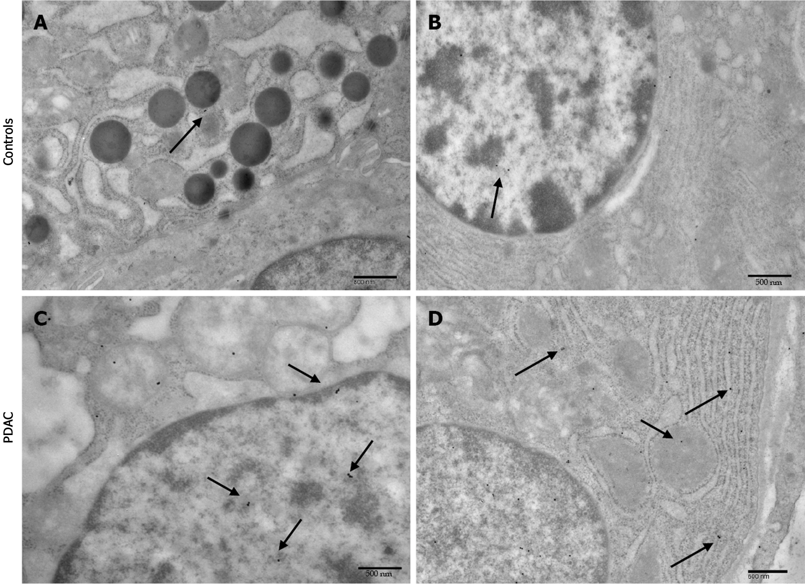
Figure 4 Prion protein immunocytochemistry in controls and pancreatic ductal adenocarcinoma.
A: Prion protein immunocytochemistry in controls. Few particles of gold prion protein (PrPc) are evident (arrows) in the cytosol (magnification: 6000 ×, scale bar: 800 nm); B: Few particles of gold PrPc are evident (arrows) in the nucleus (magnification: 8000 ×, scale bar: 500 nm); C: Prion protein immunocytochemistry in pancreatic ductal adenocarcinoma. The arrows highlight some of the PrPc immuno-gold particles in the nucleus (magnification: 8000 ×, scale bar: 500 nm); D: The arrows highlight some of the PrPc immuno-gold particles in the cytosol (magnification: 7000 ×, scale bar: 600 nm).
- Citation: Bianchini M, Giambelluca MA, Scavuzzo MC, Di Franco G, Guadagni S, Palmeri M, Furbetta N, Gianardi D, Funel N, Ricci C, Gaeta R, Pollina LE, Falcone A, Vivaldi C, Di Candio G, Biagioni F, Busceti CL, Morelli L, Fornai F. Detailing the ultrastructure’s increase of prion protein in pancreatic adenocarcinoma. World J Gastroenterol 2021; 27(42): 7324-7339
- URL: https://www.wjgnet.com/1007-9327/full/v27/i42/7324.htm
- DOI: https://dx.doi.org/10.3748/wjg.v27.i42.7324


