Copyright
©The Author(s) 2021.
World J Gastroenterol. Nov 14, 2021; 27(42): 7324-7339
Published online Nov 14, 2021. doi: 10.3748/wjg.v27.i42.7324
Published online Nov 14, 2021. doi: 10.3748/wjg.v27.i42.7324
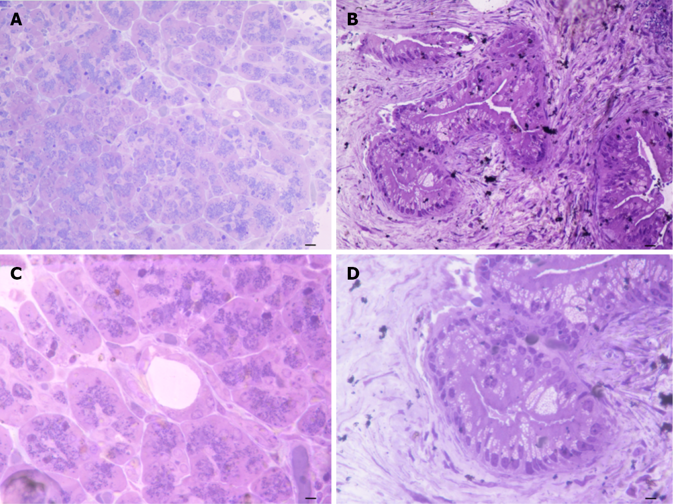
Figure 2 Semi-thin sections of control and pancreatic ductal adenocarcinoma pancreas.
A: Pancreas from control tissue at low magnification (magnification: 20 ×, scale bar: 12.5 μm). The ductal areas are evident as empty roundish areas surrounded by cell staining as much as those in the neighboring parenchyma; B: The normal pancreatic tissue characteristics are more evident at higher magnification, where there is a pale toluidine staining of ductal cells (magnification: 40 ×, scale bar: 6.25 μm); C: In pancreatic ductal adenocarcinoma (PDAC) tissue a highly non-homogeneous tissue is present and ductal regions are markedly stained (magnification: 20 ×, scale bar: 12.5 μm); D: PDAC tissue at higher magnification (magnification: 40 ×, scale bar: 6.25 μm). The ductal cells are overwhelmed and they tend to occlude the ductal lumen.
- Citation: Bianchini M, Giambelluca MA, Scavuzzo MC, Di Franco G, Guadagni S, Palmeri M, Furbetta N, Gianardi D, Funel N, Ricci C, Gaeta R, Pollina LE, Falcone A, Vivaldi C, Di Candio G, Biagioni F, Busceti CL, Morelli L, Fornai F. Detailing the ultrastructure’s increase of prion protein in pancreatic adenocarcinoma. World J Gastroenterol 2021; 27(42): 7324-7339
- URL: https://www.wjgnet.com/1007-9327/full/v27/i42/7324.htm
- DOI: https://dx.doi.org/10.3748/wjg.v27.i42.7324


