Copyright
©The Author(s) 2021.
World J Gastroenterol. Oct 28, 2021; 27(40): 6939-6950
Published online Oct 28, 2021. doi: 10.3748/wjg.v27.i40.6939
Published online Oct 28, 2021. doi: 10.3748/wjg.v27.i40.6939
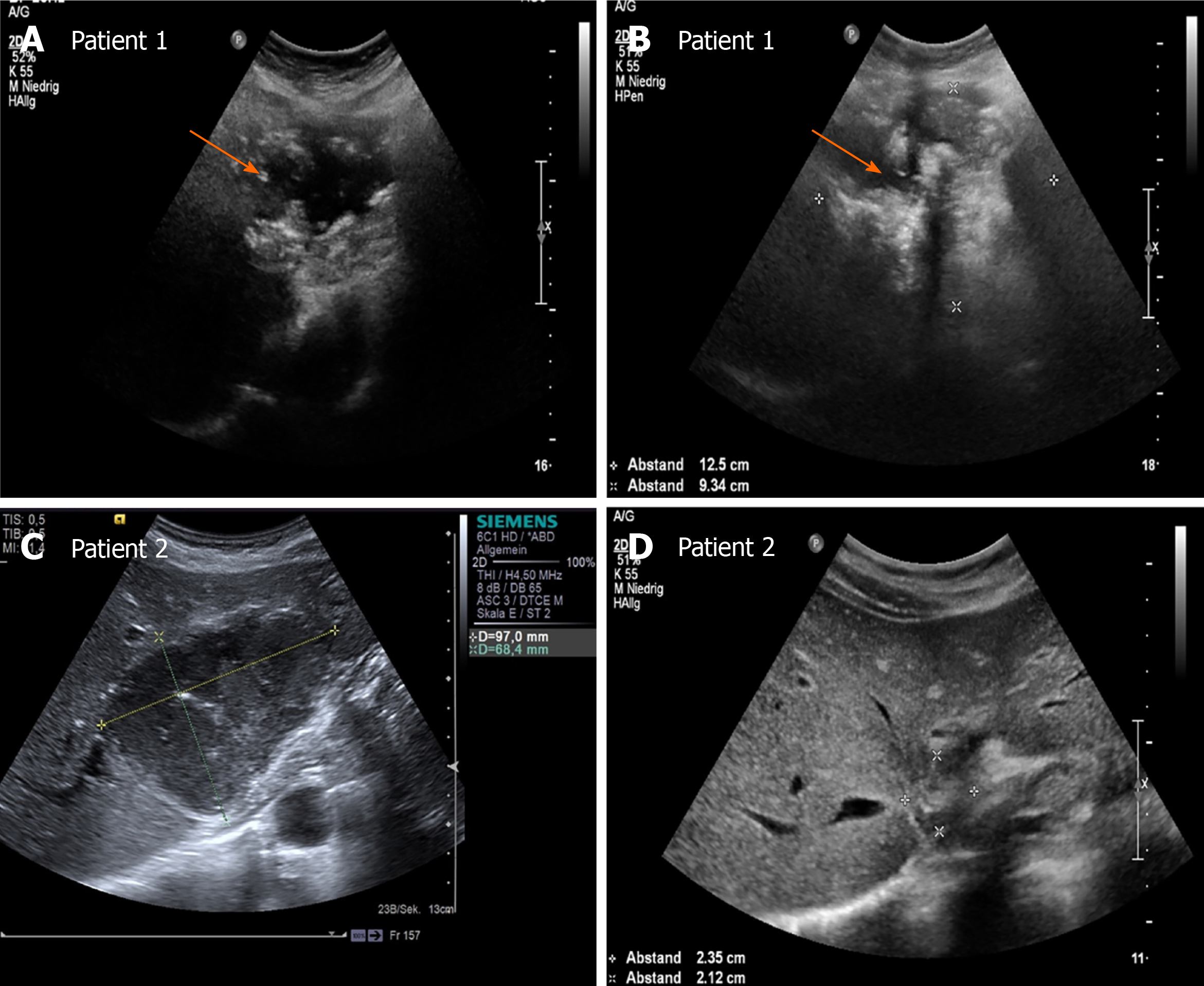
Figure 5 Hepatic alveolar echinococcosis reference lesions (arrow) in segment II/IVa/VIII (patient 1) and segment II/III (patient 2), respectively.
A and C: initially classified as pseudocystic pattern (patient 1: A, patient 2: C); B and D: In the course of each case loss of the centrally echo-poor, liquid-impressive zone and increasing calcification structures, indicating a change in sonomorphology with a transition to a hailstorm pattern (patient 1: B; patient 2: D). The ultrasound images shown are from the current patient collective.
- Citation: Schuhbaur J, Schweizer M, Philipp J, Schmidberger J, Schlingeloff P, Kratzer W. Long-term follow-up of liver alveolar echinococcosis using echinococcosis multilocularis ultrasound classification. World J Gastroenterol 2021; 27(40): 6939-6950
- URL: https://www.wjgnet.com/1007-9327/full/v27/i40/6939.htm
- DOI: https://dx.doi.org/10.3748/wjg.v27.i40.6939


