Copyright
©The Author(s) 2021.
World J Gastroenterol. Oct 28, 2021; 27(40): 6939-6950
Published online Oct 28, 2021. doi: 10.3748/wjg.v27.i40.6939
Published online Oct 28, 2021. doi: 10.3748/wjg.v27.i40.6939
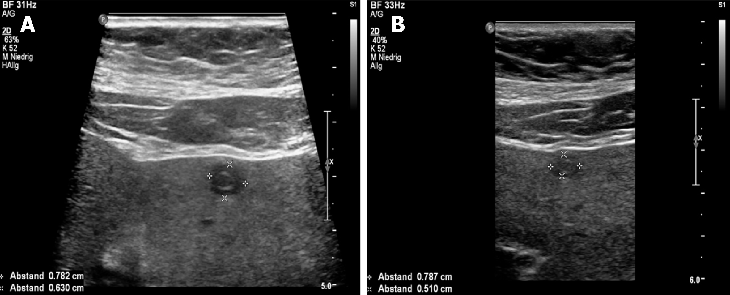
Figure 4 Hepatic alveolar echinococcosis reference lesion in segment II with pseudocystic pattern.
A: Initially sharply delineated with individual centrally echo-rich portions; B: Through the disease course, more blurred delineation and increase of centrally echo-rich portions. The ultrasound images shown are from the current patient collective.
- Citation: Schuhbaur J, Schweizer M, Philipp J, Schmidberger J, Schlingeloff P, Kratzer W. Long-term follow-up of liver alveolar echinococcosis using echinococcosis multilocularis ultrasound classification. World J Gastroenterol 2021; 27(40): 6939-6950
- URL: https://www.wjgnet.com/1007-9327/full/v27/i40/6939.htm
- DOI: https://dx.doi.org/10.3748/wjg.v27.i40.6939


