Copyright
©The Author(s) 2021.
World J Gastroenterol. Jan 28, 2021; 27(4): 371-376
Published online Jan 28, 2021. doi: 10.3748/wjg.v27.i4.371
Published online Jan 28, 2021. doi: 10.3748/wjg.v27.i4.371
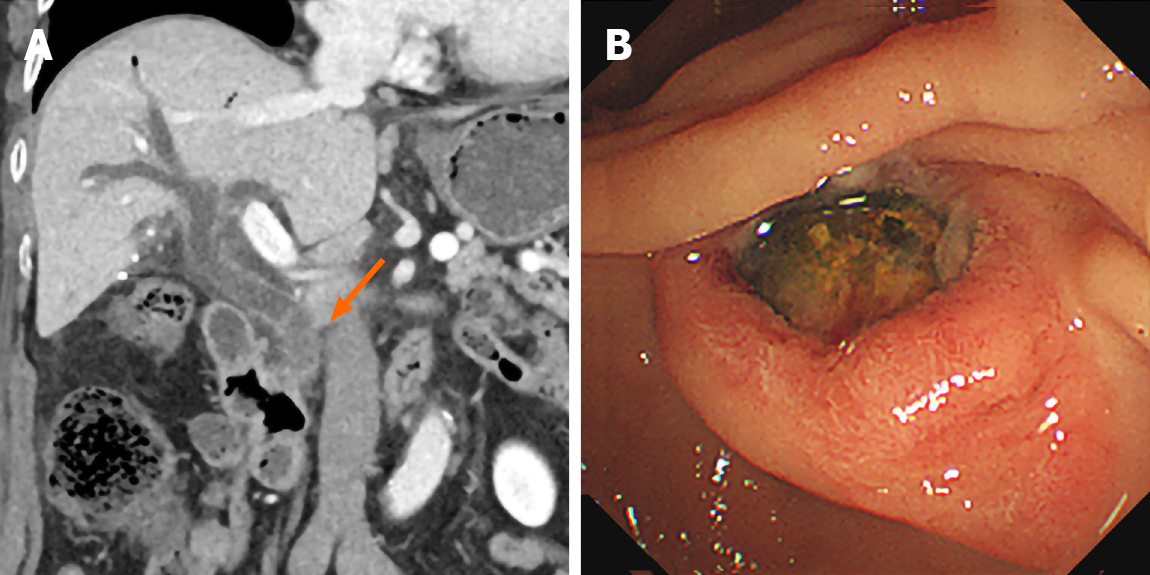
Figure 2 Computed tomography imaging and endoscopic retrograde cholangiopancreatography.
A: Follow-up computed tomography demonstrated choledocholithiasis in the extrahepatic bile duct draining the right lobe of the liver; B: An impacted stone was identified at the ampulla of Vater.
- Citation: Hwang JS, Ko SW. Duplication of the common bile duct manifesting as recurrent pyogenic cholangitis: A case report. World J Gastroenterol 2021; 27(4): 371-376
- URL: https://www.wjgnet.com/1007-9327/full/v27/i4/371.htm
- DOI: https://dx.doi.org/10.3748/wjg.v27.i4.371


