Copyright
©The Author(s) 2021.
World J Gastroenterol. Oct 21, 2021; 27(39): 6615-6630
Published online Oct 21, 2021. doi: 10.3748/wjg.v27.i39.6615
Published online Oct 21, 2021. doi: 10.3748/wjg.v27.i39.6615
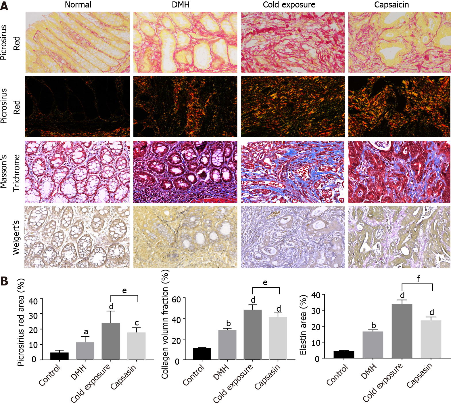
Figure 3 Changes in extracellular matrix components (collagen fibers and elastin) in colonic mucosa of different treatment groups.
A: Representative photographs of colonic tissues in rats of normal, 1,2-dimethylhyrazine (DMH), cold exposure and capsaicin groups using Masson’s trichrome: collagen (blue), nuclei and cytoplasm (red); picrosirius red in bright-field: collagen (red); polarized light: collagen (yellow-orange to green birefringence) and Weigert’s Resorcin-Fuschin: elastin (blue-black), myofibers (yellow). Magnification, × 400, scalar bar 20 μm; B: Quantitative analysis of picrosirius red staining, trichrome and Weigert’s staining as a measure of collagen and elastin density.
- Citation: Qin JC, Yu WT, Li HX, Liang YQ, Nong FF, Wen B. Cold exposure and capsaicin promote 1,2-dimethylhyrazine-induced colon carcinogenesis in rats correlates with extracellular matrix remodeling. World J Gastroenterol 2021; 27(39): 6615-6630
- URL: https://www.wjgnet.com/1007-9327/full/v27/i39/6615.htm
- DOI: https://dx.doi.org/10.3748/wjg.v27.i39.6615


