Copyright
©The Author(s) 2021.
World J Gastroenterol. Oct 21, 2021; 27(39): 6615-6630
Published online Oct 21, 2021. doi: 10.3748/wjg.v27.i39.6615
Published online Oct 21, 2021. doi: 10.3748/wjg.v27.i39.6615
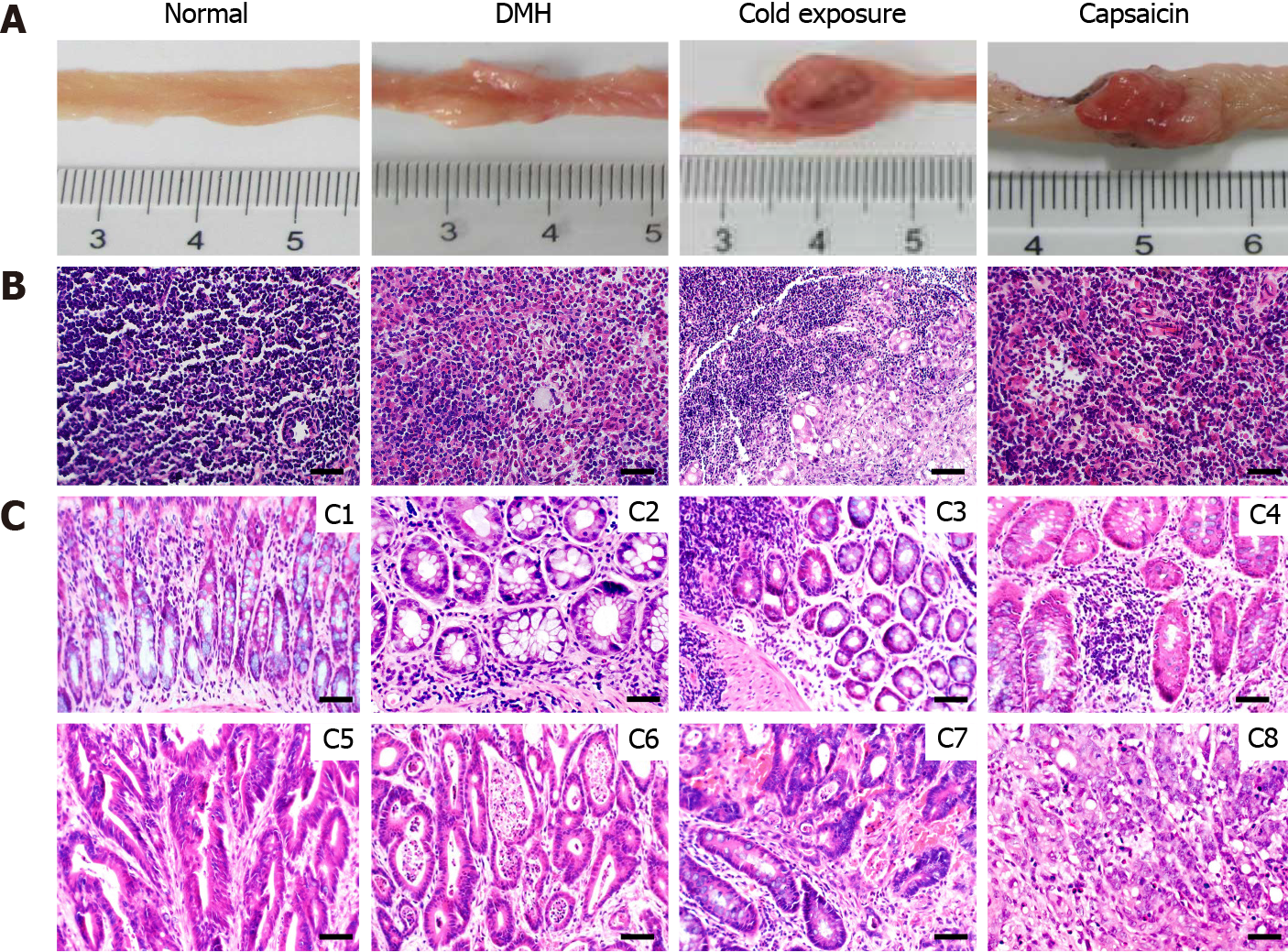
Figure 2 Pathological observation and lymph node metastases of different groups.
A: Macroscopic image of the colonic tumors; B: Representative sections stained with hematoxylin and eosin (HE) showing the histopathology of the mesocolic lymph node in the different groups; C: Representative sections stained with HE showing the histopathology of the colonic mucosa in the different groups. Normal architecture of colon was observed in the control groups (C1), adenoma with mild dysplasia with massive infiltration of inflammatory cells (C2). Histology of adenoma with moderate dysplasia in cold exposure groups (C3). Histology of adenoma with severe dysplasia (C4). Histology of well-differentiated tubular adenocarcinomas (C5). Histology of Moderately differentiated adenocarcinomas (C6). Histology of Poorly differentiated adenocarcinomas (C7). Histology of Mucinous adenocarcinoma with signet ring cells (C8) (HE staining, × 400, scalar bar 20 μm). DMH: 1,2-Dimethylhyrazine.
- Citation: Qin JC, Yu WT, Li HX, Liang YQ, Nong FF, Wen B. Cold exposure and capsaicin promote 1,2-dimethylhyrazine-induced colon carcinogenesis in rats correlates with extracellular matrix remodeling. World J Gastroenterol 2021; 27(39): 6615-6630
- URL: https://www.wjgnet.com/1007-9327/full/v27/i39/6615.htm
- DOI: https://dx.doi.org/10.3748/wjg.v27.i39.6615


