Copyright
©The Author(s) 2021.
World J Gastroenterol. Oct 14, 2021; 27(38): 6357-6373
Published online Oct 14, 2021. doi: 10.3748/wjg.v27.i38.6357
Published online Oct 14, 2021. doi: 10.3748/wjg.v27.i38.6357
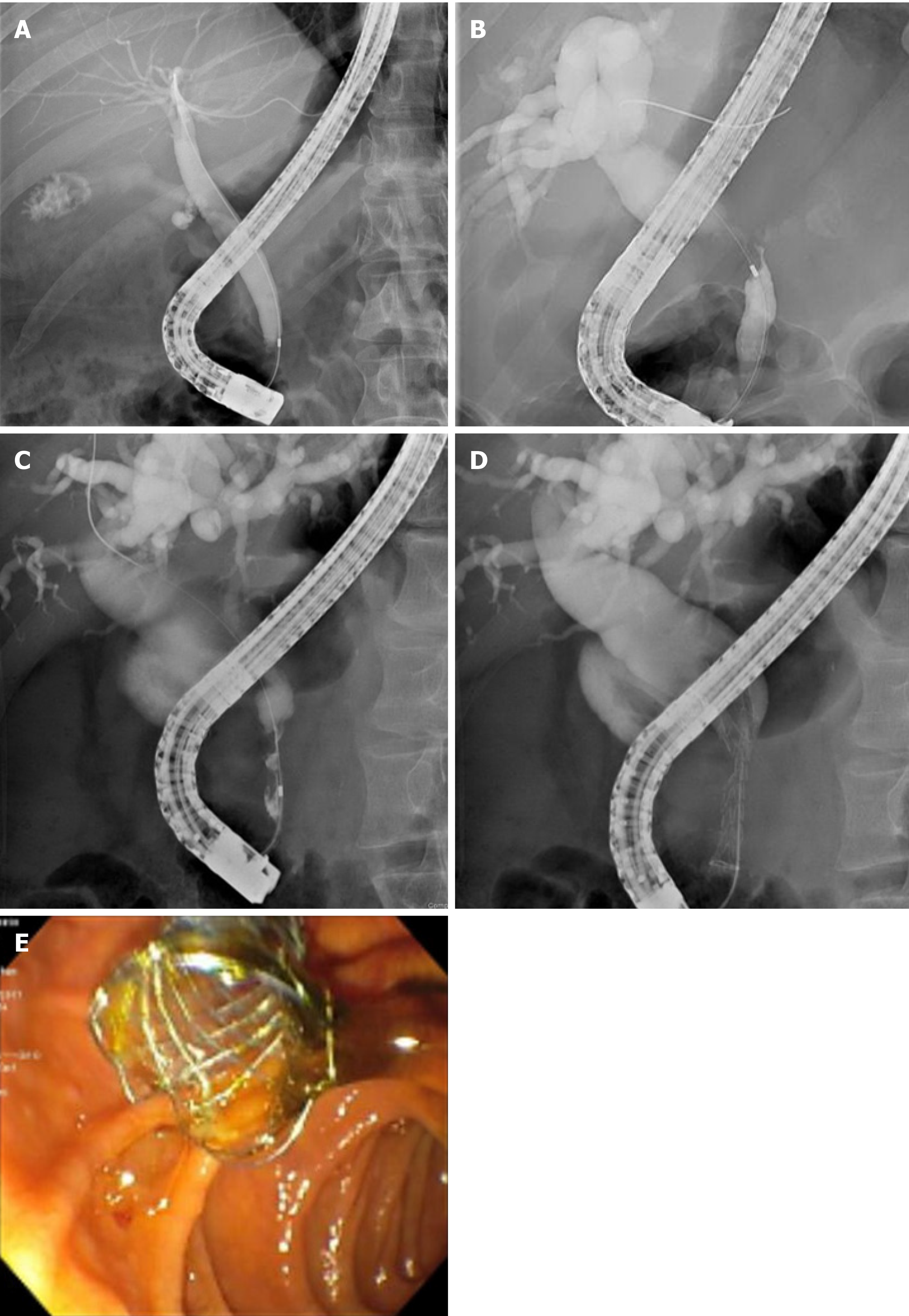
Figure 2 Endoscopic retrograde cholangiopancreatography showing cholangiocarcinoma in mid and distal common bile duct compared to normal anatomy.
A: Normal anatomy demomonstrating patent cystic, common bile, and intrahepatic ducts; B: Mid common bile duct biliary stricture with dilated common bile duct and intrahepatic ducts; C: Distal common bile duct biliary stricture with dilated common bile duct and intrahepaptic ducts; D: Fully covered self-expandable metal stent (FCSEMS) placement in the common bile duct; E: Endoscopic view of FCSEMS placed in the distal common bile duct.
- Citation: Lam R, Muniraj T. Fully covered metal biliary stents: A review of the literature. World J Gastroenterol 2021; 27(38): 6357-6373
- URL: https://www.wjgnet.com/1007-9327/full/v27/i38/6357.htm
- DOI: https://dx.doi.org/10.3748/wjg.v27.i38.6357


