Copyright
©The Author(s) 2021.
World J Gastroenterol. Oct 7, 2021; 27(37): 6290-6305
Published online Oct 7, 2021. doi: 10.3748/wjg.v27.i37.6290
Published online Oct 7, 2021. doi: 10.3748/wjg.v27.i37.6290
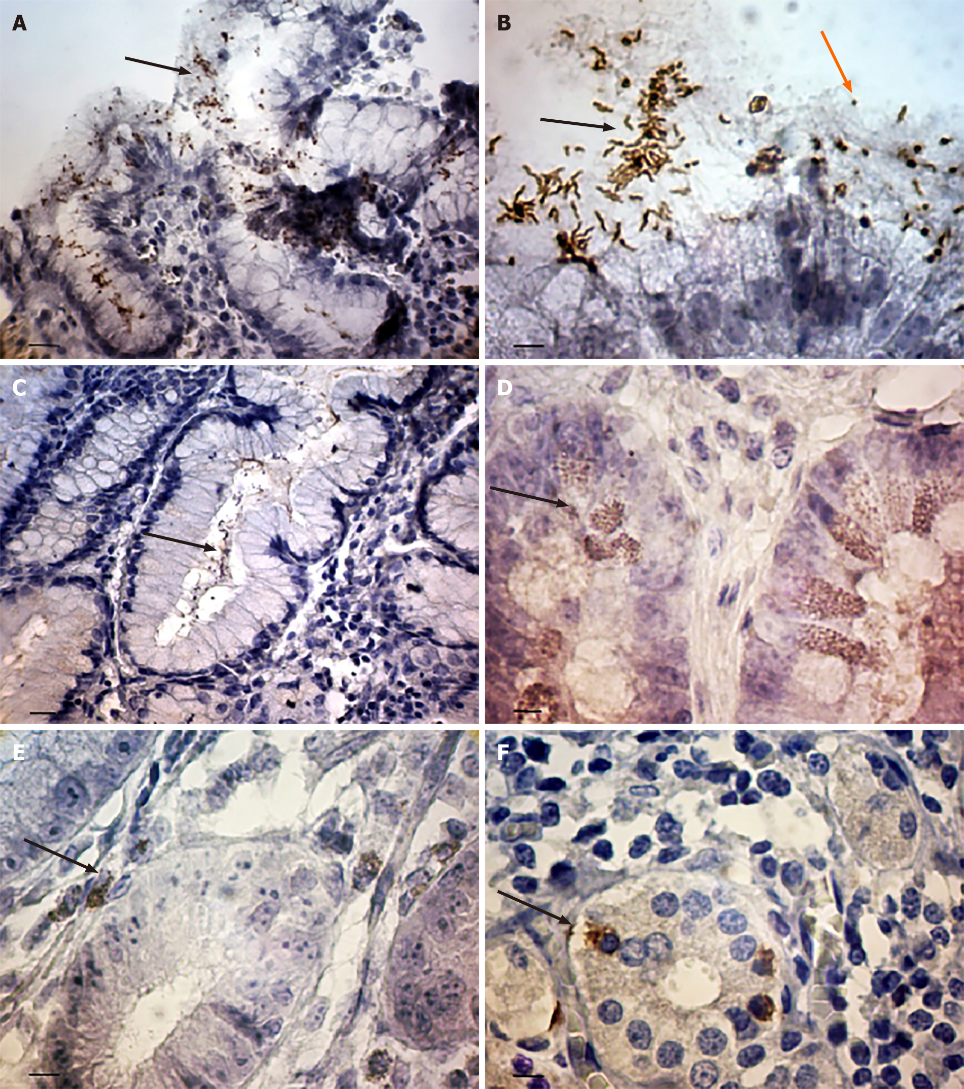
Figure 1 The features of Helicobacter pylori localization in gastric mucosa in patients with gastric cancer.
A: Coccoid forms of Helicobacter pylori (H. pylori) in the gastric pit. The some bacteria within the cytoplasm of epithelium cells (arrows); B: The spiral (black arrows) and coccoid (orange arrows) forms of H. pylori on the surface of superficial-foveolar gastric epithelium; C: The bacteria in the surface mucous gel layer of stomach (arrows); D: The point inclusions giving a positive reaction with antibodies to H. pylori within the cytoplasm of epithelial cells of deep gastric glands (arrows); E: The point inclusions giving a positive reaction with antibodies to H. pylori within the cytoplasm of the immune cells of the lamina propria of gastric mucosa (arrows); F: The point inclusions giving a positive reaction with antibodies to H. pylori within the cytoplasm of intraepithelial lymphocytes (arrows). Immunoperoxidase staining with anti-H. pylori antibody, immersion. Bars: A: 20 μm; B: 10 μm; C: 20 μm; D-F: 10 μm.
- Citation: Senchukova MA, Tomchuk O, Shurygina EI. Helicobacter pylori in gastric cancer: Features of infection and their correlations with long-term results of treatment. World J Gastroenterol 2021; 27(37): 6290-6305
- URL: https://www.wjgnet.com/1007-9327/full/v27/i37/6290.htm
- DOI: https://dx.doi.org/10.3748/wjg.v27.i37.6290


