Copyright
©The Author(s) 2021.
World J Gastroenterol. Oct 7, 2021; 27(37): 6248-6261
Published online Oct 7, 2021. doi: 10.3748/wjg.v27.i37.6248
Published online Oct 7, 2021. doi: 10.3748/wjg.v27.i37.6248
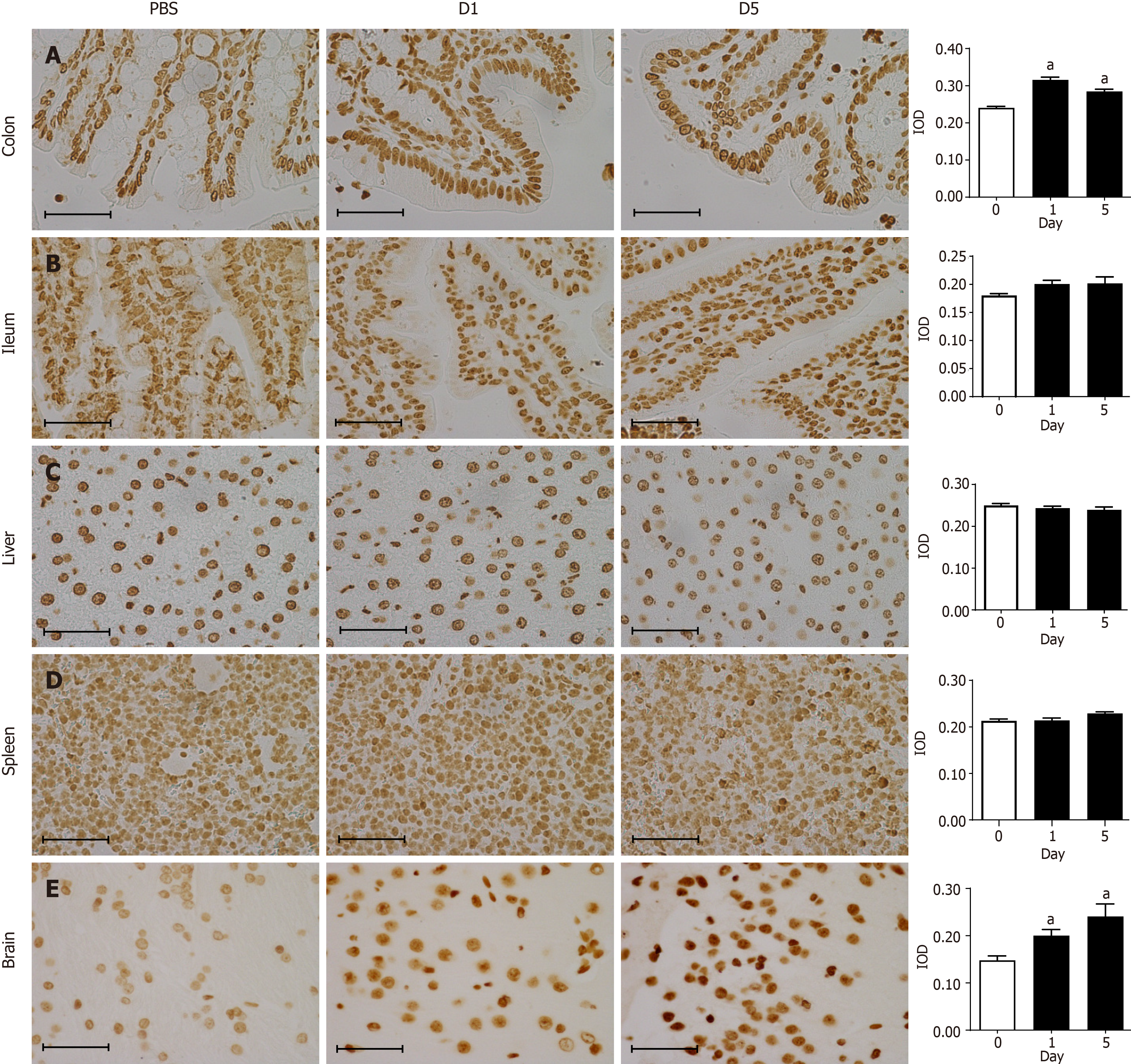
Figure 5 Immunohistochemical staining for 8-oxo-7,8-dihydro-2-deoxyguanosine in tissues.
A: Representative images showing immuno
- Citation: Nie JJ, Pian YY, Hu JH, Fan GQ, Zeng LT, Ouyang QG, Gao ZX, Liu Z, Wang CC, Liu Q, Cai JP. Increased systemic RNA oxidative damage and diagnostic value of RNA oxidative metabolites during Shigella flexneri-induced intestinal infection. World J Gastroenterol 2021; 27(37): 6248-6261
- URL: https://www.wjgnet.com/1007-9327/full/v27/i37/6248.htm
- DOI: https://dx.doi.org/10.3748/wjg.v27.i37.6248


