Copyright
©The Author(s) 2021.
World J Gastroenterol. Oct 7, 2021; 27(37): 6248-6261
Published online Oct 7, 2021. doi: 10.3748/wjg.v27.i37.6248
Published online Oct 7, 2021. doi: 10.3748/wjg.v27.i37.6248
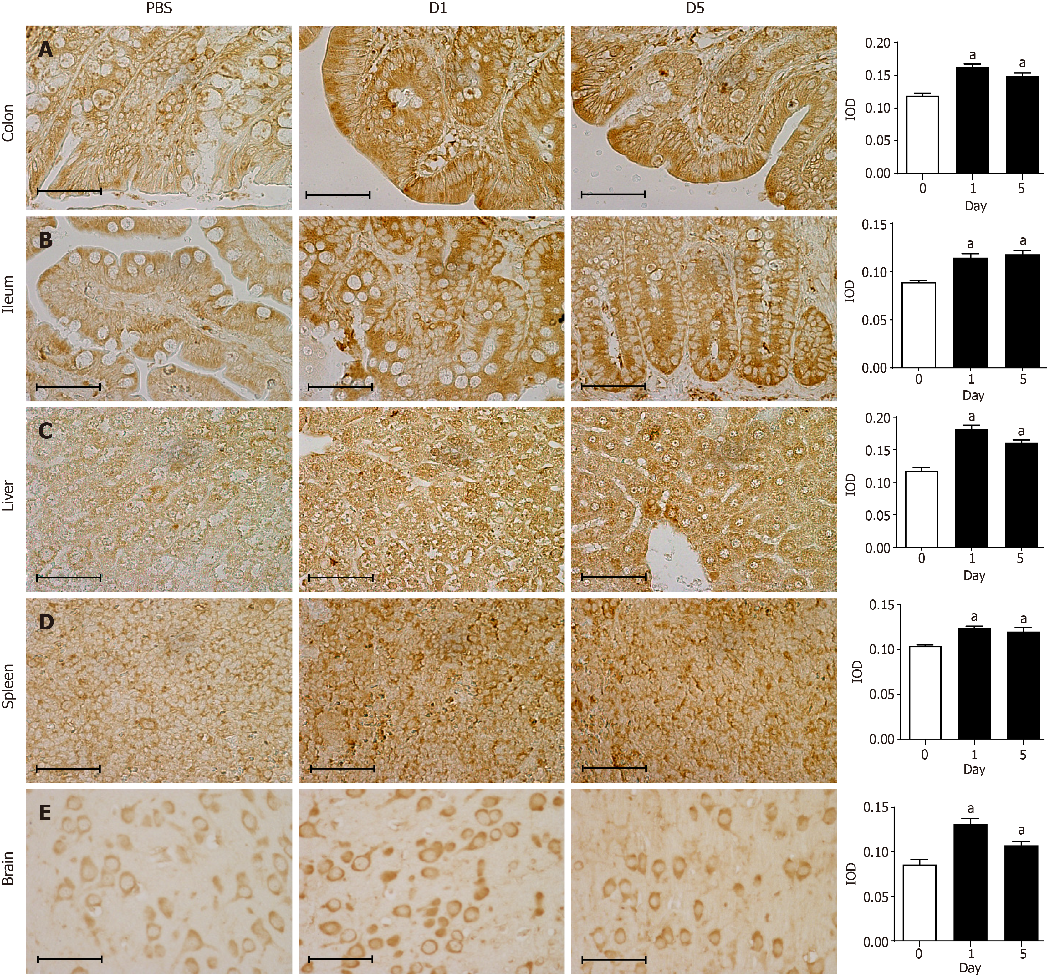
Figure 4 Immunohistochemical staining for 8-oxo-7,8-dihydroguanosine in tissues.
A: Representative images showing immunohistochemical staining (400 × magnification) of 8-oxo-7,8-dihydroguanosine (8-oxo-Gsn) in paraffin-embedded colon sections from rats. Left panel images (PBS) indicate the results of the control group. Middle panel images (D1) indicate the results one day after Shigella flexneri (S. flexneri) infection. Right panel images (D5) indicate the results five days after S. flexneri infection; B: Immunohistochemical staining for 8-oxo-Gsn in ileum sections; C: Immunohistochemical staining for 8-oxo-Gsn in liver sections; D: Immunohistochemical staining for 8-oxo-Gsn in spleen sections; E: Immunohistochemical staining for 8-oxo-Gsn in brain sections. Histogram: The quantitative analysis of immunohistochemical staining of 8-oxo-Gsn in tissues of the infection and control groups. Scale bar, 50 μm. aP < 0.05. The immunohistochemical images were analyzed using the Image Pro-Plus (IPP) software. The integrated optical density (IOD) is a representative parameter for assessing the immunostaining quantification in the IPP analyses, and it indicates the total amount of staining material in that area. IOD: Integrated optical density.
- Citation: Nie JJ, Pian YY, Hu JH, Fan GQ, Zeng LT, Ouyang QG, Gao ZX, Liu Z, Wang CC, Liu Q, Cai JP. Increased systemic RNA oxidative damage and diagnostic value of RNA oxidative metabolites during Shigella flexneri-induced intestinal infection. World J Gastroenterol 2021; 27(37): 6248-6261
- URL: https://www.wjgnet.com/1007-9327/full/v27/i37/6248.htm
- DOI: https://dx.doi.org/10.3748/wjg.v27.i37.6248


