Copyright
©The Author(s) 2021.
World J Gastroenterol. Sep 28, 2021; 27(36): 5989-6003
Published online Sep 28, 2021. doi: 10.3748/wjg.v27.i36.5989
Published online Sep 28, 2021. doi: 10.3748/wjg.v27.i36.5989
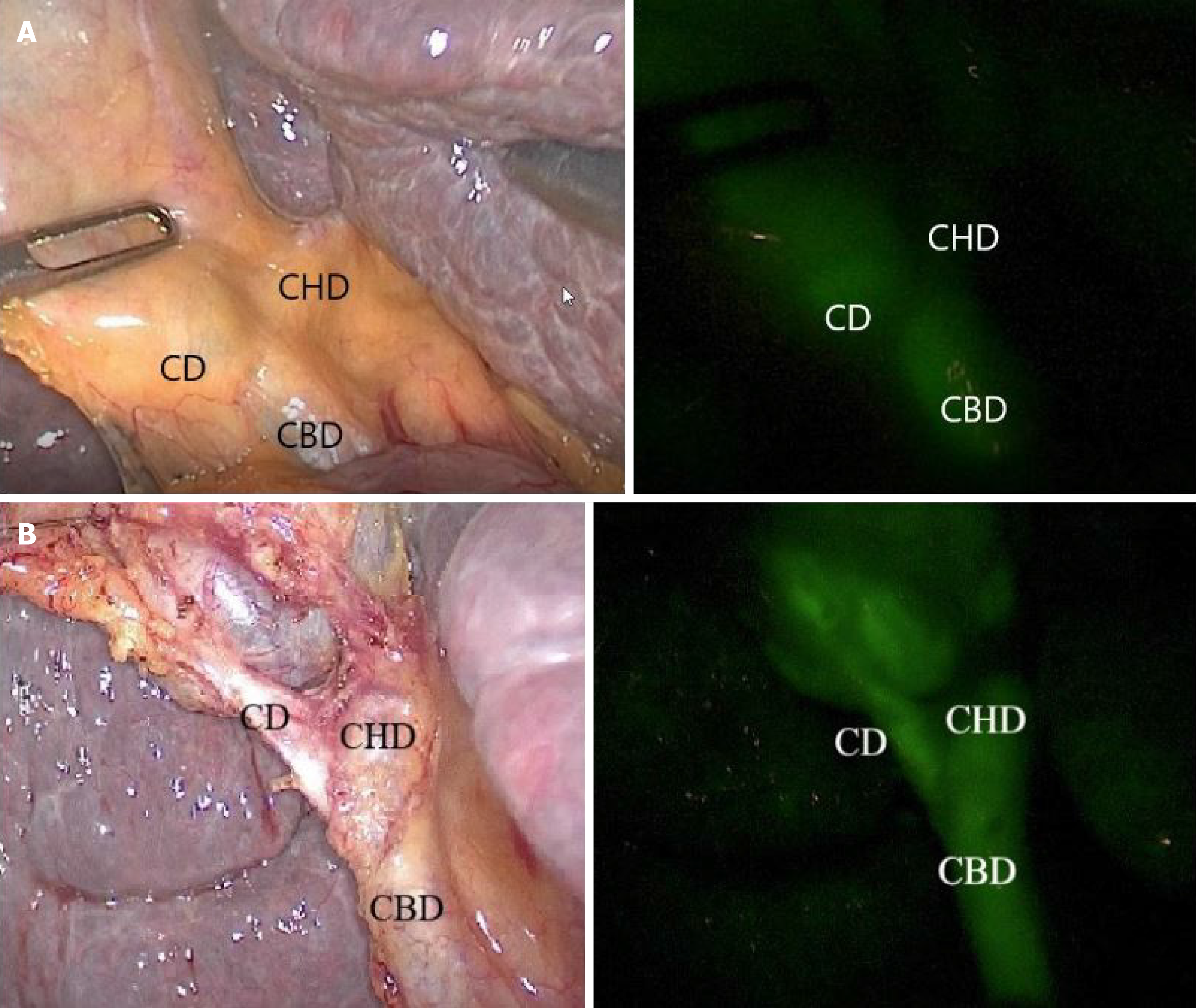
Figure 1 Intra-operative real-time identification of biliary structures in a cirrhotic patient, with visible light to the left and NIRF-C to the right.
A: Pre-dissection visualization of biliary anatomy; B: After complete dissection. One can observe the posterior implantation of the cystic duct on the common hepatic duct. CD: Cystic duct; CHD: Common hepatic duct; CBD: Common bile duct.
- Citation: Pesce A, Piccolo G, Lecchi F, Fabbri N, Diana M, Feo CV. Fluorescent cholangiography: An up-to-date overview twelve years after the first clinical application. World J Gastroenterol 2021; 27(36): 5989-6003
- URL: https://www.wjgnet.com/1007-9327/full/v27/i36/5989.htm
- DOI: https://dx.doi.org/10.3748/wjg.v27.i36.5989


