Copyright
©The Author(s) 2021.
World J Gastroenterol. Sep 21, 2021; 27(35): 5978-5988
Published online Sep 21, 2021. doi: 10.3748/wjg.v27.i35.5978
Published online Sep 21, 2021. doi: 10.3748/wjg.v27.i35.5978
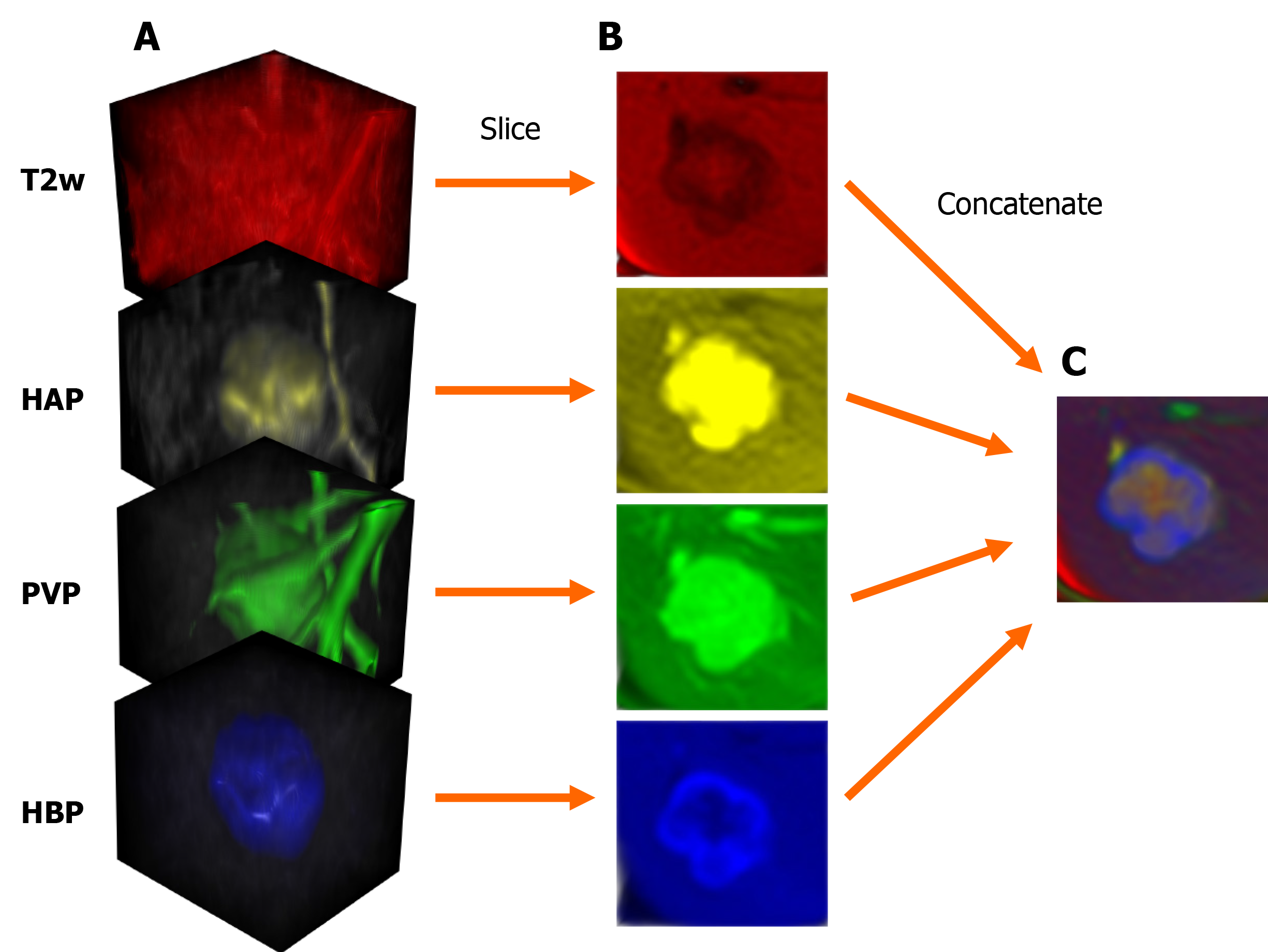
Figure 2 Steps of input data preparation for the two-dimensional densely connected convolutional neural network.
A: Cubic magnetic resonance image volumes containing the lesion; B: Axial slices acquired from the cropped volumes; C: The four axial slices are concatenated into one three-dimensional image; each slice is represented by a different color. T2w: T2-weighted; HAP: Hepatic arterial phase; PVP: Portal venous phase; HBP: Hepatobiliary phase.
- Citation: Stollmayer R, Budai BK, Tóth A, Kalina I, Hartmann E, Szoldán P, Bérczi V, Maurovich-Horvat P, Kaposi PN. Diagnosis of focal liver lesions with deep learning-based multi-channel analysis of hepatocyte-specific contrast-enhanced magnetic resonance imaging. World J Gastroenterol 2021; 27(35): 5978-5988
- URL: https://www.wjgnet.com/1007-9327/full/v27/i35/5978.htm
- DOI: https://dx.doi.org/10.3748/wjg.v27.i35.5978


