Copyright
©The Author(s) 2021.
World J Gastroenterol. Sep 7, 2021; 27(33): 5460-5473
Published online Sep 7, 2021. doi: 10.3748/wjg.v27.i33.5460
Published online Sep 7, 2021. doi: 10.3748/wjg.v27.i33.5460
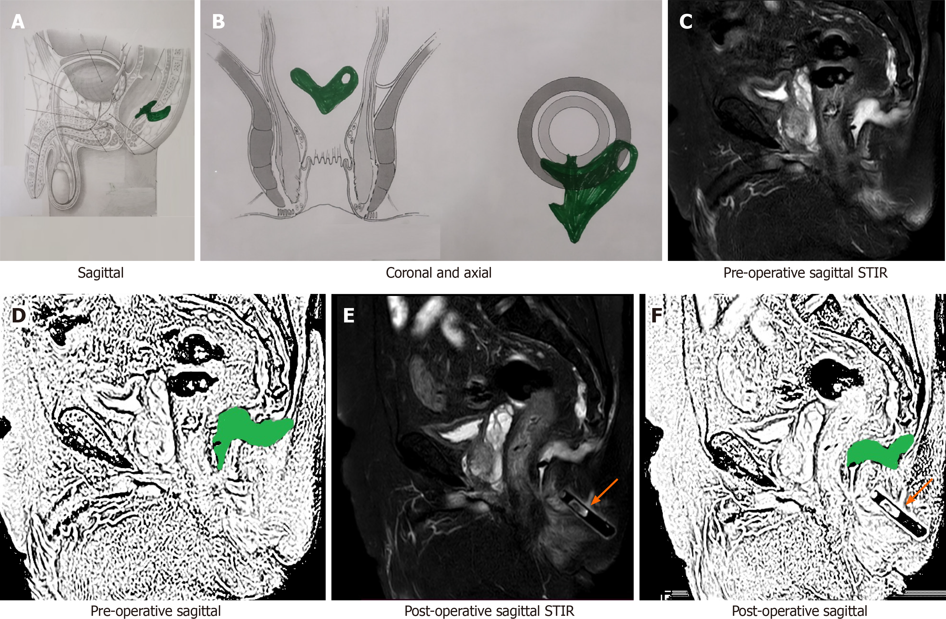
Figure 7 A 69-year-old male patient with a posterior fistula with a presacral abscess.
A wide bore drainage tube was inserted into the cavity intraoperatively. However, the patient did not improve. Postoperative check magnetic resonance imaging (MRI) demonstrated that the tube was in the wrong plane and the presacral abscess was not properly drained. A: Axial section (schematic diagram); B: Coronal section (schematic diagram); C: Preoperative short-T1 inversion recovery (STIR) sagittal MRI; D: Sketch of pre-operative sagittal MRI image highlighting anal fistula and presacral abscess (green color); E: Postoperative STIR sagittal MRI showing drainage tube in the wrong plane and persistent presacral abscess; F: Sketch of postoperative sagittal MRI highlighting persisting anal fistula and presacral abscess (green color) (orange arrows show the drainage tube). STIR: Short-T1 inversion recovery.
- Citation: Garg P, Kaur B, Yagnik VD, Dawka S, Menon GR. Guidelines on postoperative magnetic resonance imaging in patients operated for cryptoglandular anal fistula: Experience from 2404 scans. World J Gastroenterol 2021; 27(33): 5460-5473
- URL: https://www.wjgnet.com/1007-9327/full/v27/i33/5460.htm
- DOI: https://dx.doi.org/10.3748/wjg.v27.i33.5460


