Copyright
©The Author(s) 2021.
World J Gastroenterol. Sep 7, 2021; 27(33): 5460-5473
Published online Sep 7, 2021. doi: 10.3748/wjg.v27.i33.5460
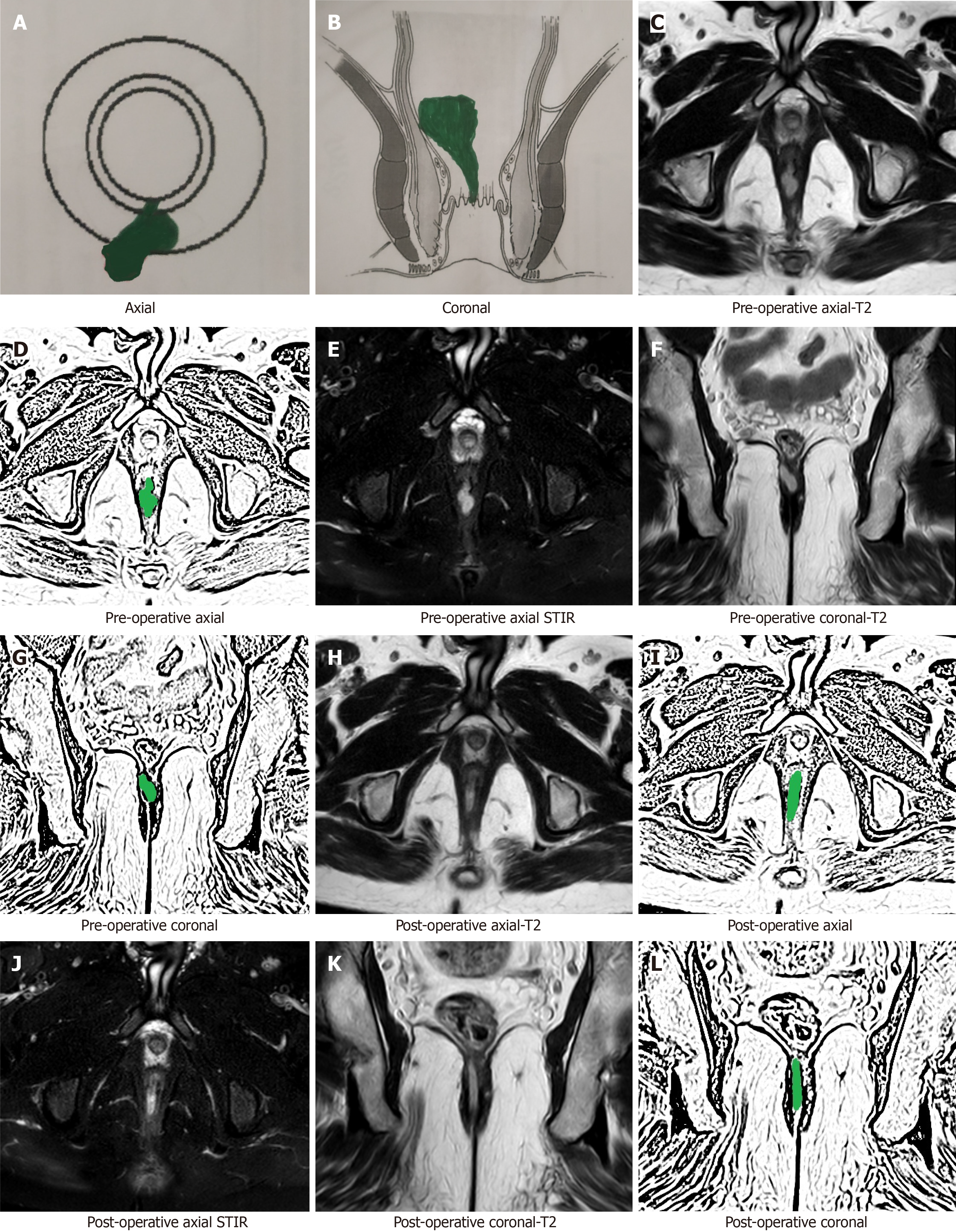
Figure 1 A 37-year-old male patient with a high posterior intersphincteric abscess was drained by transanal opening of the inter sphincteric space procedure.
Magnetic resonance imaging (MRI) done after 3 wk after surgery showed hyperintense signal (due to healing granulation tissue) at the same location similar to the hyperintense signal in the preoperative images (due to pus collection). A: Axial section (schematic diagram); B: Coronal section (schematic diagram); C: Pre-operative T2-weighted MRI; D: Sketch of pre-operative axial MRI image highlighting abscess (green color); E: Pre-operative short-T1 inversion recovery (STIR) axial MRI; F: Pre-operative T2-weighted coronal MRI; G: Sketch of pre-operative coronal MRI image highlighting abscess (green color); H: Postoperative T2-weighted axial MRI; I: Sketch of postoperative axial MRI image highlighting healing granulation tissue (green color); J: Postoperative STIR axial MRI; K: Postoperative T2-weighted coronal MRI; L: Sketch of postoperative coronal MRI image highlighting healing granulation tissue (green color). STIR: Short-T1 inversion recovery.
- Citation: Garg P, Kaur B, Yagnik VD, Dawka S, Menon GR. Guidelines on postoperative magnetic resonance imaging in patients operated for cryptoglandular anal fistula: Experience from 2404 scans. World J Gastroenterol 2021; 27(33): 5460-5473
- URL: https://www.wjgnet.com/1007-9327/full/v27/i33/5460.htm
- DOI: https://dx.doi.org/10.3748/wjg.v27.i33.5460