Copyright
©The Author(s) 2021.
World J Gastroenterol. Aug 21, 2021; 27(31): 5288-5296
Published online Aug 21, 2021. doi: 10.3748/wjg.v27.i31.5288
Published online Aug 21, 2021. doi: 10.3748/wjg.v27.i31.5288
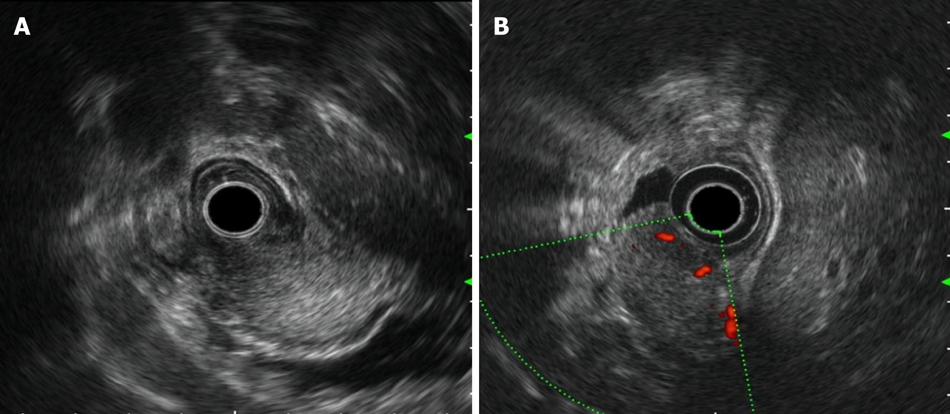
Figure 2 Rich blood flow in the lesion under Doppler ultrasound.
A: Endoscopic ultrasonography revealed that the lesion originated from the submucosal layer, which was heterogeneous and hyperechoic, and the posterior muscularis propria and serosal surface were present; B: The blood flow was abundant on Doppler ultrasound.
- Citation: Wu JD, Chen YX, Luo C, Xu FH, Zhang L, Hou XH, Song J. Plexiform angiomyxoid myofibroblastic tumor treated by endoscopic submucosal dissection: A case report and review of the literature. World J Gastroenterol 2021; 27(31): 5288-5296
- URL: https://www.wjgnet.com/1007-9327/full/v27/i31/5288.htm
- DOI: https://dx.doi.org/10.3748/wjg.v27.i31.5288


