Copyright
©The Author(s) 2021.
World J Gastroenterol. Aug 14, 2021; 27(30): 5060-5075
Published online Aug 14, 2021. doi: 10.3748/wjg.v27.i30.5060
Published online Aug 14, 2021. doi: 10.3748/wjg.v27.i30.5060
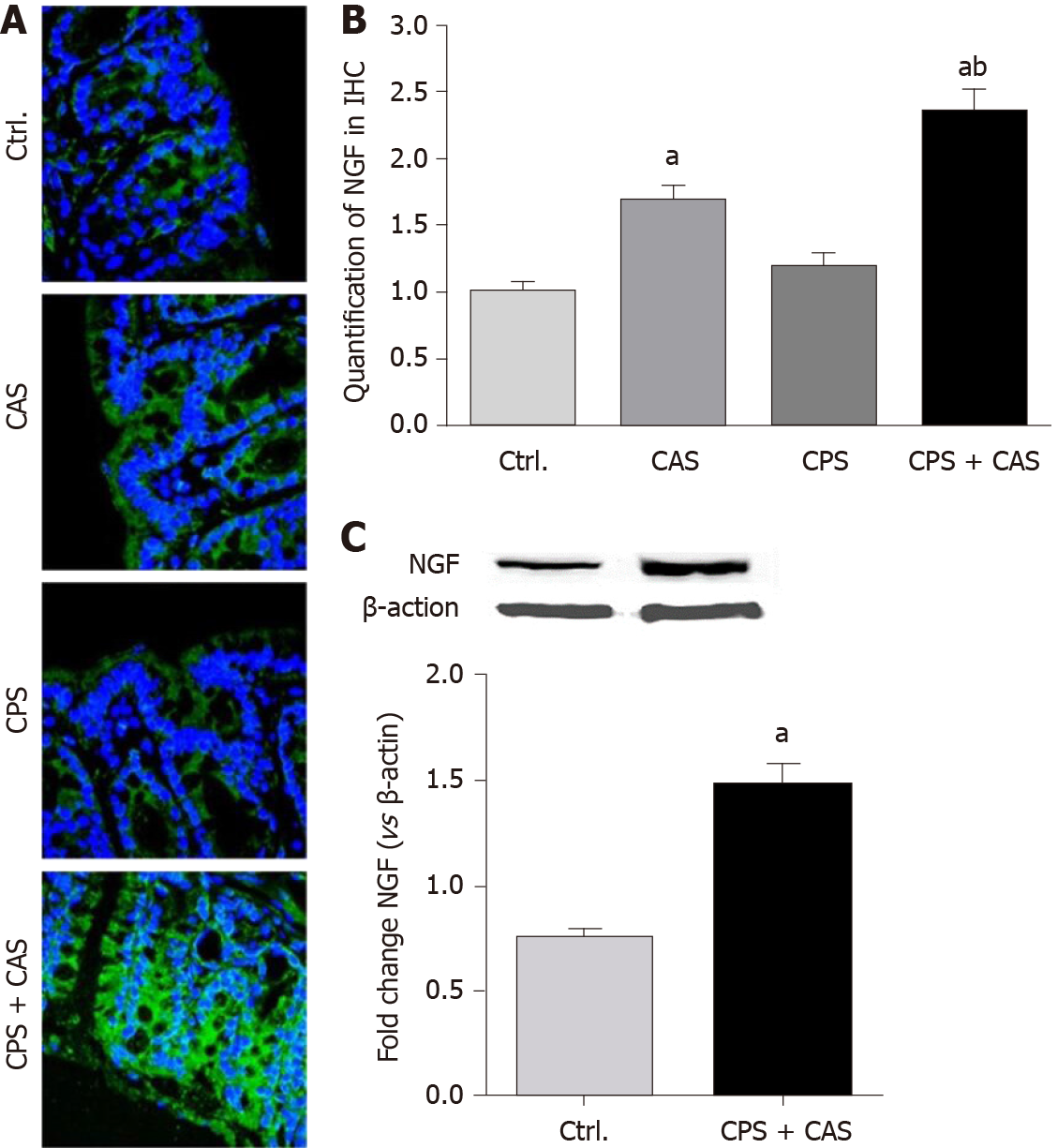
Figure 6 Nerve growth factor expression level in the colon wall.
A: Immunohistochemical staining of nerve growth factor (NGF; green) was detected with nuclear counterstaining staining (blue) in controls, chronic adult stress (CAS), chronic prenatal stress (CPS) and CPS + CAS group female rat colon walls. × 400 magnification representative pictures were shown; B: Quantification of NGF levels from colon wall in immunohistochemistry (IHC) (n = 4 rats in each group, one-way ANOVA, aP < 0.05 vs control group; bP < 0.05 vs CPS group); C: Western blots of NGF protein from control and CPS + CAS female rats colon wall tissue (n = 6 rats in each group, t-test, aP < 0.05 vs control group).
- Citation: Chen JH, Sun Y, Ju PJ, Wei JB, Li QJ, Winston JH. Estrogen augmented visceral pain and colonic neuron modulation in a double-hit model of prenatal and adult stress. World J Gastroenterol 2021; 27(30): 5060-5075
- URL: https://www.wjgnet.com/1007-9327/full/v27/i30/5060.htm
- DOI: https://dx.doi.org/10.3748/wjg.v27.i30.5060


