Copyright
©The Author(s) 2021.
World J Gastroenterol. Aug 7, 2021; 27(29): 4862-4878
Published online Aug 7, 2021. doi: 10.3748/wjg.v27.i29.4862
Published online Aug 7, 2021. doi: 10.3748/wjg.v27.i29.4862
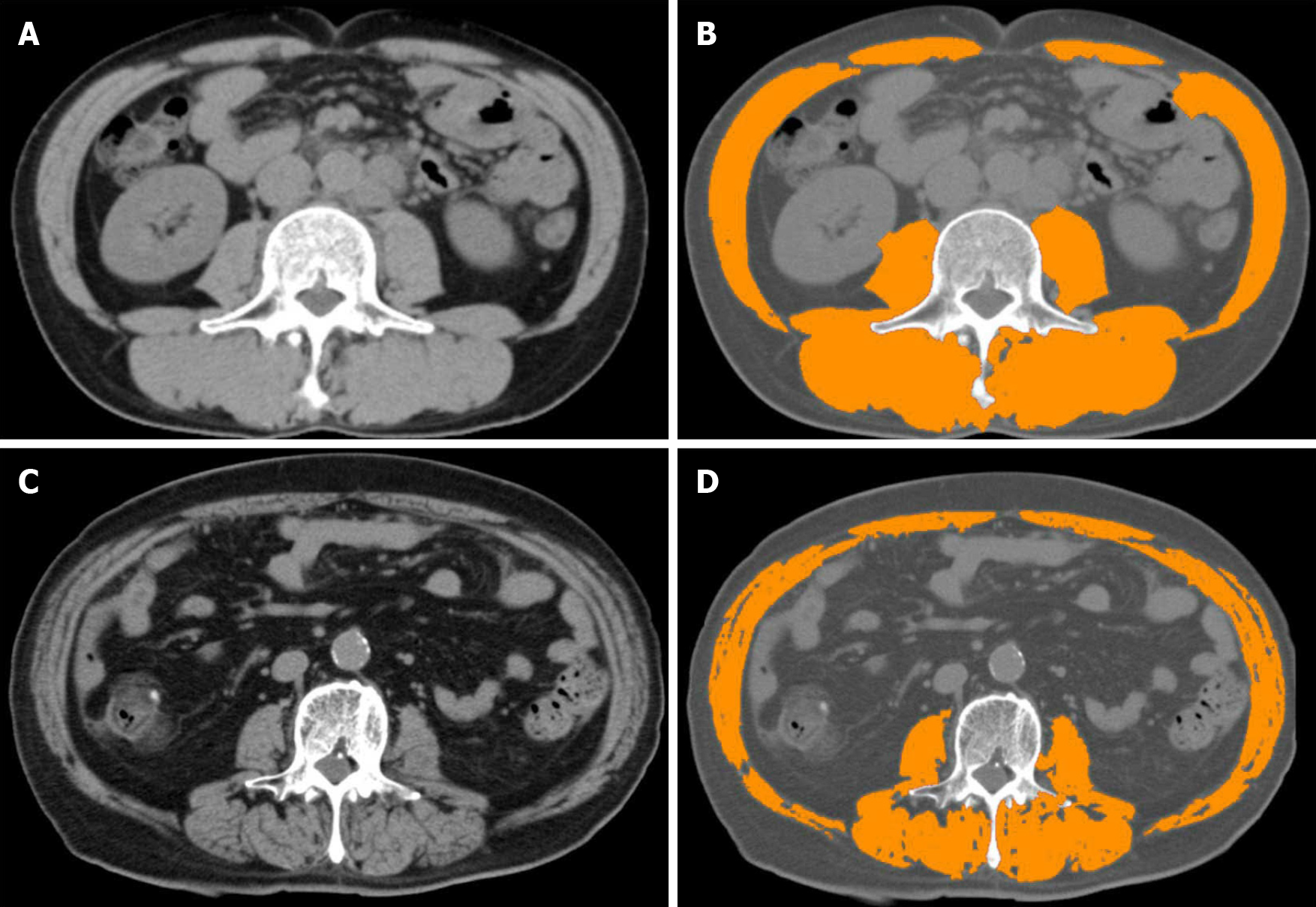
Figure 3 Computed tomography modality and skeletal muscle index.
A and C: Analysing computed tomography slices obtained at the third lumbar vertebra level; B: Patient with normal skeletal muscle index (SMI) values; D: Patient with reduced SMI values. After using medical imaging software and analysing areas of predefined Hounsfield units, SMI values are calculated.
- Citation: Hari A. Muscular abnormalities in liver cirrhosis. World J Gastroenterol 2021; 27(29): 4862-4878
- URL: https://www.wjgnet.com/1007-9327/full/v27/i29/4862.htm
- DOI: https://dx.doi.org/10.3748/wjg.v27.i29.4862


