Copyright
©The Author(s) 2021.
World J Gastroenterol. Jul 28, 2021; 27(28): 4738-4745
Published online Jul 28, 2021. doi: 10.3748/wjg.v27.i28.4738
Published online Jul 28, 2021. doi: 10.3748/wjg.v27.i28.4738
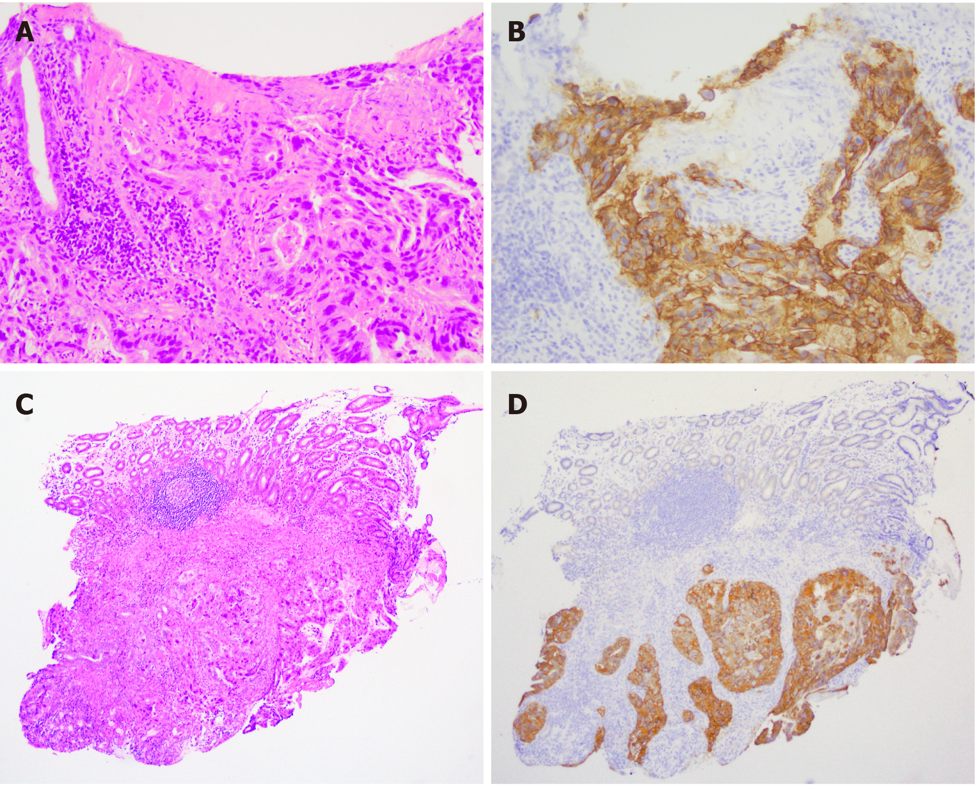
Figure 2 Histological findings of biopsy specimens from duodenal lesion.
A: Adenocarcinoma cells proliferate in the proper mucosal and submucosal layer with ulcer formation (HE staining, × 200); B: Adenocarcinoma cells show strong HER2 membranous expression (× 200); C and D: Adenocarcinoma cells proliferate in the submucosal layer with strong HER2 protein expression (× 54).
- Citation: Hirokawa YS, Iwata T, Okugawa Y, Tanaka K, Sakurai H, Watanabe M. HER2-positive adenocarcinoma arising from heterotopic pancreas tissue in the duodenum: A case report. World J Gastroenterol 2021; 27(28): 4738-4745
- URL: https://www.wjgnet.com/1007-9327/full/v27/i28/4738.htm
- DOI: https://dx.doi.org/10.3748/wjg.v27.i28.4738


