Copyright
©The Author(s) 2021.
World J Gastroenterol. Jul 28, 2021; 27(28): 4738-4745
Published online Jul 28, 2021. doi: 10.3748/wjg.v27.i28.4738
Published online Jul 28, 2021. doi: 10.3748/wjg.v27.i28.4738
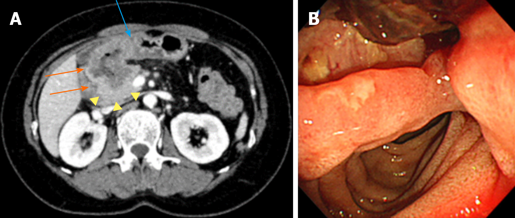
Figure 1 Abdominal computed tomography and gastroduodenoscopy.
Images were obtained before the start of treatment. A: An axial image shows an irregular circumferential mass in the first portion of duodenum (orange arrows), with transmural extension to pylorus (blue arrow) and direct extension to pancreas head (yellow arrow heads); B: During gastroduodenoscopy, mass lesion with ulcer was visible in the first portion of duodenum.
- Citation: Hirokawa YS, Iwata T, Okugawa Y, Tanaka K, Sakurai H, Watanabe M. HER2-positive adenocarcinoma arising from heterotopic pancreas tissue in the duodenum: A case report. World J Gastroenterol 2021; 27(28): 4738-4745
- URL: https://www.wjgnet.com/1007-9327/full/v27/i28/4738.htm
- DOI: https://dx.doi.org/10.3748/wjg.v27.i28.4738


