Copyright
©The Author(s) 2021.
World J Gastroenterol. Jul 21, 2021; 27(27): 4322-4341
Published online Jul 21, 2021. doi: 10.3748/wjg.v27.i27.4322
Published online Jul 21, 2021. doi: 10.3748/wjg.v27.i27.4322
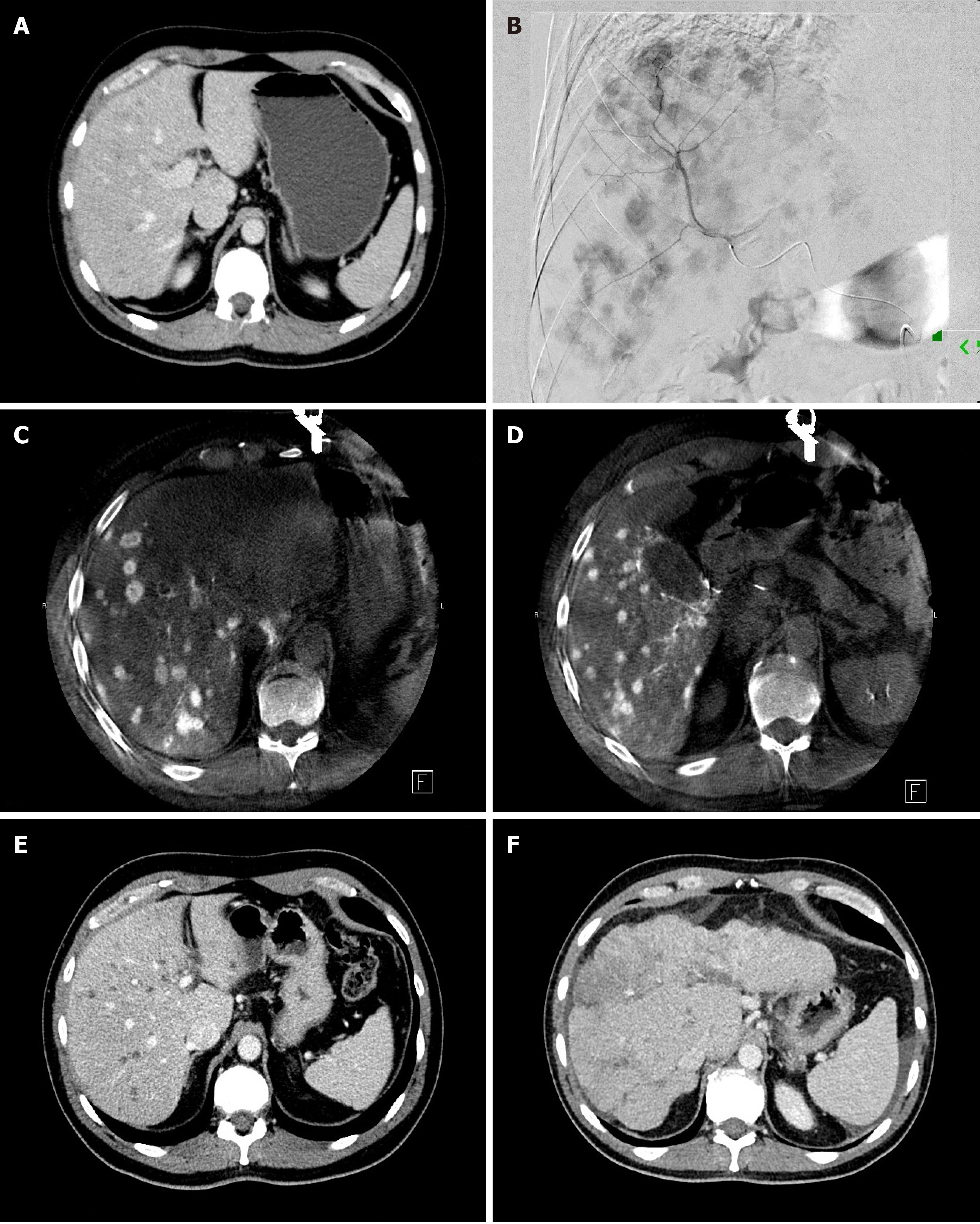
Figure 4 Transarterial radioembolization of pancreatic adenocarcinoma liver metastases.
A: Pre-treatment contrast-enhanced computed tomography (CT) image of a 45-year-old man with pancreatic cancer demonstrates bilobar multifocal mildly hypodense liver metastases; B: Hepatic angiogram with contrast injection into the anterior division of the right hepatic artery shows multifocal arterially enhancing liver metastases; C and D: Intra-procedural cone-beam CT images of the liver during selective contrast injection into right hepatic artery shows multifocal arterially enhancing liver metastases; E: Contrast-enhanced CT image 1 mo after bilobar transarterial radioembolization (TARE) shows hypodense, “burnt out” liver metastases. The patient CA19-9 Level decreased from 882 U/mL pre-procedure to 56 U/mL. One year after the initial TARE new liver lesions appeared and his CA19-9 increased to 530 U/mL. He underwent a second round of bilobar TARE, and his CA19-9 Level decreased to 233 U/mL; F: Contrast-enhanced CT image 18 mo after the initial TARE shows shrunken liver with lobulated, nodular borders consistent with TARE-induced pseudocirrhosis. The patient died 22 mo after the initial TARE due to extrahepatic and hepatic progression of his disease.
- Citation: Bibok A, Kim DW, Malafa M, Kis B. Minimally invasive image-guided therapy of primary and metastatic pancreatic cancer. World J Gastroenterol 2021; 27(27): 4322-4341
- URL: https://www.wjgnet.com/1007-9327/full/v27/i27/4322.htm
- DOI: https://dx.doi.org/10.3748/wjg.v27.i27.4322


